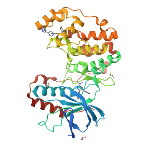
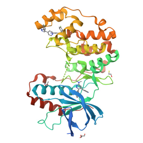
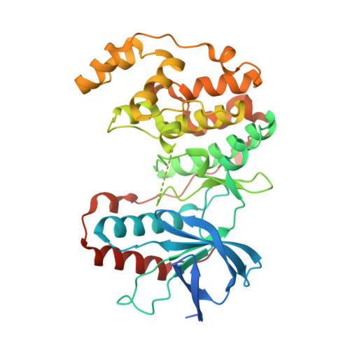
4F9W

Human P38alpha MAPK in Complex with a Novel and Selective Small Molecule Inhibitor


Experimental Data Snapshot

  • Method: X-RAY DIFFRACTION
  • Resolution: 2.00 Å
  • R-Value Free: 0.216 
  • R-Value Work: 0.171 
  • R-Value Observed: 0.173 

Starting Model: experimental
View more details

wwPDB Validation   3D Report Full Report


Ligand Structure Quality Assessment 


This is version 2.1 of the entry. See complete history


Literature

Development of Novel In Vivo Chemical Probes to Address CNS Protein Kinase Involvement in Synaptic Dysfunction.

Watterson, D.M.Grum-Tokars, V.L.Roy, S.M.Schavocky, J.P.Bradaric, B.D.Bachstetter, A.D.Xing, B.Dimayuga, E.Saeed, F.Zhang, H.Staniszewski, A.Pelletier, J.C.Minasov, G.Anderson, W.F.Arancio, O.Van Eldik, L.J.

(2013) PLoS One 8: e66226-e66226

  • DOI: https://doi.org/10.1371/journal.pone.0066226
  • Primary Citation of Related Structures:  
    4F9W, 4F9Y, 4FA2

  • PubMed Abstract: 

    Serine-threonine protein kinases are critical to CNS function, yet there is a dearth of highly selective, CNS-active kinase inhibitors for in vivo investigations. Further, prevailing assumptions raise concerns about whether single kinase inhibitors can show in vivo efficacy for CNS pathologies, and debates over viable approaches to the development of safe and efficacious kinase inhibitors are unsettled. It is critical, therefore, that these scientific challenges be addressed in order to test hypotheses about protein kinases in neuropathology progression and the potential for in vivo modulation of their catalytic activity. Identification of molecular targets whose in vivo modulation can attenuate synaptic dysfunction would provide a foundation for future disease-modifying therapeutic development as well as insight into cellular mechanisms. Clinical and preclinical studies suggest a critical link between synaptic dysfunction in neurodegenerative disorders and the activation of p38αMAPK mediated signaling cascades. Activation in both neurons and glia also offers the unusual potential to generate enhanced responses through targeting a single kinase in two distinct cell types involved in pathology progression. However, target validation has been limited by lack of highly selective inhibitors amenable to in vivo use in the CNS. Therefore, we employed high-resolution co-crystallography and pharmacoinformatics to design and develop a novel synthetic, active site targeted, CNS-active, p38αMAPK inhibitor (MW108). Selectivity was demonstrated by large-scale kinome screens, functional GPCR agonist and antagonist analyses of off-target potential, and evaluation of cellular target engagement. In vitro and in vivo assays demonstrated that MW108 ameliorates beta-amyloid induced synaptic and cognitive dysfunction. A serendipitous discovery during co-crystallographic analyses revised prevailing models about active site targeting of inhibitors, providing insights that will facilitate future kinase inhibitor design. Overall, our studies deliver highly selective in vivo probes appropriate for CNS investigations and demonstrate that modulation of p38αMAPK activity can attenuate synaptic dysfunction.


  • Organizational Affiliation

    Department of Molecular Pharmacology and Biological Chemistry, Northwestern University, Chicago, Illinois, United States of America.


Macromolecules
Find similar proteins by:  (by identity cutoff)  |  3D Structure
Entity ID: 1
MoleculeChains Sequence LengthOrganismDetailsImage
Mitogen-activated protein kinase 14383Homo sapiensMutation(s): 0 
Gene Names: MAPK14CSBPCSBP1CSBP2CSPB1MXI2SAPK2A
EC: 2.7.11.24
UniProt & NIH Common Fund Data Resources
Find proteins for Q16539 (Homo sapiens)
Explore Q16539 
Go to UniProtKB:  Q16539
PHAROS:  Q16539
GTEx:  ENSG00000112062 
Entity Groups  
Sequence Clusters30% Identity50% Identity70% Identity90% Identity95% Identity100% Identity
UniProt GroupQ16539
Sequence Annotations
Expand
  • Reference Sequence
Small Molecules
Ligands 6 Unique
IDChains Name / Formula / InChI Key2D Diagram3D Interactions
LM4
Query on LM4

Download Ideal Coordinates CCD File 
D [auth A]N,N-dimethyl-6-(naphthalen-2-yl)-5-(pyridin-4-yl)pyridazin-3-amine
C21 H18 N4
ODFGOBPLQKLCRM-UHFFFAOYSA-N
GG5
Query on GG5

Download Ideal Coordinates CCD File 
B [auth A],
C [auth A]
4-[3-(4-FLUOROPHENYL)-1H-PYRAZOL-4-YL]PYRIDINE
C14 H10 F N3
BILJSHVAAVZERY-UHFFFAOYSA-N
GOL
Query on GOL

Download Ideal Coordinates CCD File 
H [auth A]GLYCEROL
C3 H8 O3
PEDCQBHIVMGVHV-UHFFFAOYSA-N
BME
Query on BME

Download Ideal Coordinates CCD File 
G [auth A]BETA-MERCAPTOETHANOL
C2 H6 O S
DGVVWUTYPXICAM-UHFFFAOYSA-N
ZN
Query on ZN

Download Ideal Coordinates CCD File 
E [auth A]ZINC ION
Zn
PTFCDOFLOPIGGS-UHFFFAOYSA-N
ACT
Query on ACT

Download Ideal Coordinates CCD File 
F [auth A]ACETATE ION
C2 H3 O2
QTBSBXVTEAMEQO-UHFFFAOYSA-M
Binding Affinity Annotations 
IDSourceBinding Affinity
LM4 PDBBind:  4F9W Ki: 114 (nM) from 1 assay(s)
Experimental Data & Validation

Experimental Data

  • Method: X-RAY DIFFRACTION
  • Resolution: 2.00 Å
  • R-Value Free: 0.216 
  • R-Value Work: 0.171 
  • R-Value Observed: 0.173 
  • Space Group: P 21 21 21
Unit Cell:
Length ( Å )Angle ( ˚ )
a = 65.671α = 90
b = 74.311β = 90
c = 77.27γ = 90
Software Package:
Software NamePurpose
HKL-2000data collection
PHASERphasing
REFMACrefinement
HKL-2000data reduction
HKL-2000data scaling

Structure Validation

View Full Validation Report


Ligand Structure Quality Assessment 


Entry History & Funding Information

Deposition Data


Funding OrganizationLocationGrant Number
Alzheimerss Drug Discovery Foundation (ADDF)United States261108
National Institutes of Health/National Institute on Aging (NIH/NIA)United StatesU01AG043415
National Institutes of Health/National Institute on Aging (NIH/NIA)United StatesR01AG031311
National Institutes of Health/National Institute of Neurological Disorders and Stroke (NIH/NINDS)United StatesR01NS056051
National Institutes of Health/National Institute of Neurological Disorders and Stroke (NIH/NINDS)United StatesR01NS093920

Revision History  (Full details and data files)

  • Version 1.0: 2013-06-05
    Type: Initial release
  • Version 1.1: 2013-06-12
    Changes: Database references
  • Version 1.2: 2013-07-24
    Changes: Database references
  • Version 2.0: 2020-01-01
    Changes: Author supporting evidence, Database references, Polymer sequence
  • Version 2.1: 2023-09-13
    Changes: Data collection, Database references, Derived calculations, Refinement description