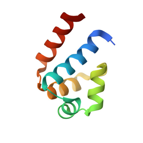
Structural and Mutagenic Analysis of the RM Controller Protein C.Esp1396I.
Martin, R.N., McGeehan, J.E., Kneale, G.(2014) PLoS One 9: e98365-e98365
- PubMed: 24887147
- DOI: https://doi.org/10.1371/journal.pone.0098365
- Primary Citation of Related Structures:
4F8D, 4FBI, 4FN3, 4I6R, 4I6T, 4I6U, 4IA8, 4IVZ - PubMed Abstract:
Bacterial restriction-modification (RM) systems are comprised of two complementary enzymatic activities that prevent the establishment of foreign DNA in a bacterial cell: DNA methylation and DNA restriction. These two activities are tightly regulated to prevent over-methylation or auto-restriction. Many Type II RM systems employ a controller (C) protein as a transcriptional regulator for the endonuclease gene (and in some cases, the methyltransferase gene also). All high-resolution structures of C-protein/DNA-protein complexes solved to date relate to C.Esp1396I, from which the interactions of specific amino acid residues with DNA bases and/or the phosphate backbone could be observed. Here we present both structural and DNA binding data for a series of mutations to the key DNA binding residues of C.Esp1396I. Our results indicate that mutations to the backbone binding residues (Y37, S52) had a lesser affect on DNA binding affinity than mutations to those residues that bind directly to the bases (T36, R46), and the contributions of each side chain to the binding energies are compared. High-resolution X-ray crystal structures of the mutant and native proteins showed that the fold of the proteins was unaffected by the mutations, but also revealed variation in the flexible loop conformations associated with DNA sequence recognition. Since the tyrosine residue Y37 contributes to DNA bending in the native complex, we have solved the structure of the Y37F mutant protein/DNA complex by X-ray crystallography to allow us to directly compare the structure of the DNA in the mutant and native complexes.
- Biophysics Laboratories, School of Biological Sciences, Institute of Biomedical and Biomolecular Science, University of Portsmouth, Portsmouth, United Kingdom.
Organizational Affiliation:


