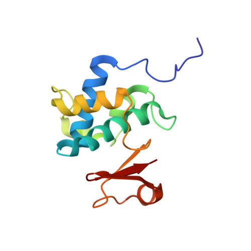
4F55 | pdb_00004f55

Crystal Structure of the Catalytic Domain of the Bacillus cereus SleB Protein


Experimental Data Snapshot

  • Method: X-RAY DIFFRACTION
  • Resolution: 1.85 Å
  • R-Value Free: 
    0.212 (Depositor), 0.231 (DCC) 
  • R-Value Work: 
    0.180 (Depositor), 0.193 (DCC) 
  • R-Value Observed: 
    0.181 (Depositor) 

wwPDB Validation 3D Report Full Report

Validation slider image for 4F55

This is version 1.3 of the entry. See complete history. 

Re-refinement Note

A newer entry is available that reflects an alternative modeling of the original data: 6TCI

Literature

Crystal Structure of the Catalytic Domain of the Bacillus cereus SleB Protein, Important in Cortex Peptidoglycan Degradation during Spore Germination.

Li, Y., Jin, K., Setlow, B., Setlow, P., Hao, B.

(2012) J Bacteriol 194: 4537-4545

  • DOI: https://doi.org/10.1128/JB.00877-12
  • Primary Citation Related Structures: 
    4F55

  • PubMed Abstract: 

    The SleB protein is one of two redundant cortex-lytic enzymes (CLEs) that initiate the degradation of cortex peptidoglycan (PG), a process essential for germination of spores of Bacillus species, including Bacillus anthracis. SleB has been characterized as a soluble lytic transglycosylase that specifically recognizes spore cortex PG and catalyzes the cleavage of glycosidic bonds between N-acetylmuramic acid (NAM) and N-acetylglucosamine residues with concomitant formation of a 1,6-anhydro bond in the NAM residue. We found that like the full-length Bacillus cereus SleB, the catalytic C-terminal domain (SleB(C)) exhibited high degradative activity on cortex PG in vitro, although SleB's N-terminal domain, thought to bind PG, was inactive. The 1.85-Å crystal structure of SleB(C) reveals an ellipsoid molecule with two distinct domains dominated by either α helices or β strands. The overall fold of SleB closely resembles that of the catalytic domain of the family 1 lytic transglycosylases but with a completely different topological arrangement. Structural analysis shows that an invariant Glu157 of SleB is in a position equivalent to that of the catalytic glutamate in other lytic transglycosylases. Indeed, SleB bearing a Glu157-to-Gln mutation lost its cortex degradative activity completely. In addition, the other redundant CLE (called CwlJ) in Bacillus species likely has a three-dimensional structure similar to that of SleB, including the invariant putative catalytic Glu residue. SleB and CwlJ may offer novel targets for the development of anti-spore agents.


  • Organizational Affiliation: 
    • Department of Molecular, Microbial and Structural Biology University of Connecticut Health Center, Farmington, Connecticut, USA.

Macromolecule Content 

  • Total Structure Weight: 14.19 kDa 
  • Atom Count: 1,232 
  • Modeled Residue Count: 128 
  • Deposited Residue Count: 128 
  • Unique protein chains: 1

Macromolecules

Find similar proteins by:|  3D Structure
Entity ID: 1
MoleculeChains  Sequence LengthOrganismDetailsImage
Spore cortex-lytic enzyme128Bacillus cereus ATCC 14579Mutation(s): 0 
Gene Names: BC_2753, sleB
UniProt
Find proteins for P0A3V0 (Bacillus cereus (strain ATCC 14579 / DSM 31 / CCUG 7414 / JCM 2152 / NBRC 15305 / NCIMB 9373 / NCTC 2599 / NRRL B-3711))
Explore P0A3V0 
Go to UniProtKB:  P0A3V0
Entity Groups
Sequence Clusters30% Identity50% Identity70% Identity90% Identity95% Identity100% Identity
UniProt GroupP0A3V0
Sequence Annotations
Expand
Reference Sequence

Experimental Data & Validation

Experimental Data

  • Method: X-RAY DIFFRACTION
  • Resolution: 1.85 Å
  • R-Value Free:  0.212 (Depositor), 0.231 (DCC) 
  • R-Value Work:  0.180 (Depositor), 0.193 (DCC) 
  • R-Value Observed: 0.181 (Depositor) 
Space Group: I 4 3 2
Unit Cell:
Length ( Å )Angle ( ˚ )
a = 134.626α = 90
b = 134.626β = 90
c = 134.626γ = 90
Software Package:
Software NamePurpose
HKL-2000data collection
PHENIXmodel building
REFMACrefinement
HKL-2000data reduction
HKL-2000data scaling
PHENIXphasing

Structure Validation

View Full Validation Report


Entry History 

Deposition Data

  • Released Date: 2012-07-11 
  • Deposition Author(s): Hao, B.

Revision History  (Full details and data files)

  • Version 1.0: 2012-07-11
    Type: Initial release
  • Version 1.1: 2012-08-29
    Changes: Database references
  • Version 1.2: 2017-11-15
    Changes: Refinement description
  • Version 1.3: 2024-02-28
    Changes: Data collection, Database references, Derived calculations