4F3H | pdb_00004f3h

The structural of FimXEAL-c-di-GMP from Xanthomonas campestris


Experimental Data Snapshot

  • Method: X-RAY DIFFRACTION
  • Resolution: 2.50 Å
  • R-Value Free: 
    0.259 (Depositor), 0.277 (DCC) 
  • R-Value Work: 
    0.234 (Depositor), 0.281 (DCC) 
  • R-Value Observed: 
    0.234 (Depositor) 

Starting Model: experimental
View more details

wwPDB Validation 3D Report Full Report

Validation slider image for 4F3H

Ligand Structure Quality Assessment 


This is version 1.2 of the entry. See complete history

Literature

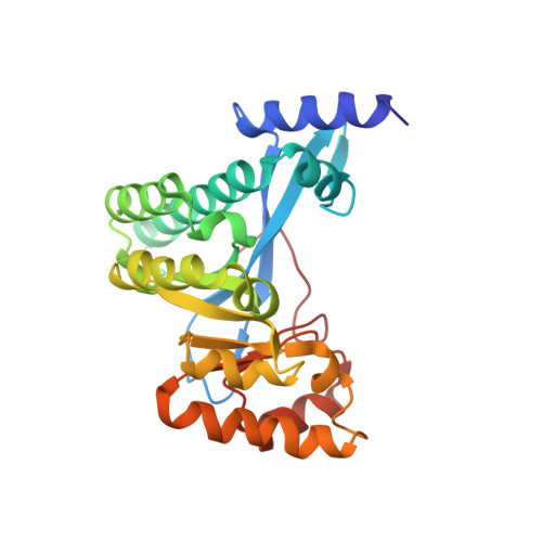
Structural polymorphism of c-di-GMP bound to an EAL domain and in complex with a type II PilZ-domain protein.

Chin, K.-H.Kuo, W.-T.Yu, Y.-J.Liao, Y.-T.Yang, M.T.Chou, S.-H.

(2012) Acta Crystallogr D Biol Crystallogr 68: 1380-1392

  • DOI: https://doi.org/10.1107/S0907444912030594
  • Primary Citation Related Structures: 
    4F3H, 4F48

  • PubMed Abstract: 

    Cyclic di-GMP (c-di-GMP) is a novel secondary-messenger molecule that is involved in regulating a plethora of important bacterial activities through binding to an unprecedented array of effectors. Proteins with a canonical PilZ domain that bind c-di-GMP play crucial roles in regulating flagellum-based motility. In contrast, noncanonical type II PilZ domains that do not effectively bind c-di-GMP regulate twitching motility, which is dependent on type IV pili (T4P). Recent data indicate that T4P biogenesis is initiated via the interaction of a noncanonical type II PilZ protein with the GGDEF/EAL-domain protein FimX and the pilus motor protein PilB at high c-di-GMP concentrations. However, the molecular details of such interactions remain to be elucidated. In this manuscript, the first hetero-complex crystal structure between a type II PilZ protein and the EAL domain of the FimX protein (FimX(EAL)) from Xanthomonas campestris pv. campestris (Xcc) in the presence of c-di-GMP is reported. This work reveals two novel conformations of monomeric c-di-GMP in the XccFimX(EAL)-c-di-GMP and XccFimX(EAL)-c-di-GMP-XccPilZ complexes, as well as a unique interaction mode of a type II PilZ domain with FimX(EAL). These findings indicate that c-di-GMP is sufficiently flexible to adjust its conformation to match the corresponding recognition motifs of different cognate effectors. Together, these results represent a first step towards an understanding of how T4P biogenesis is controlled by c-di-GMP at the molecular level and also of the ability of c-di-GMP to bind to a wide variety of effectors.


  • Organizational Affiliation
    • Agricultural Biotechnology Center, National Chung Hsing University, Taichung 40227, Taiwan.

Macromolecule Content 

  • Total Structure Weight: 28.77 kDa 
  • Atom Count: 2,178 
  • Modeled Residue Count: 247 
  • Deposited Residue Count: 250 
  • Unique protein chains: 1

Macromolecules

Find similar proteins by:|  3D Structure
Entity ID: 1
MoleculeChains  Sequence LengthOrganismDetailsImage
Putative uncharacterized protein250Xanthomonas campestris pv. campestrisMutation(s): 0 
Gene Names: XCC2291
UniProt
Find proteins for Q8P8F1 (Xanthomonas campestris pv. campestris (strain ATCC 33913 / DSM 3586 / NCPPB 528 / LMG 568 / P 25))
Explore Q8P8F1 
Go to UniProtKB:  Q8P8F1
Entity Groups
Sequence Clusters30% Identity50% Identity70% Identity90% Identity95% Identity100% Identity
UniProt GroupQ8P8F1
Sequence Annotations
Expand
Reference Sequence

Small Molecules

Ligands 1 Unique
IDChains Name / Formula / InChI Key2D Diagram3D Interactions
C2E

Query on C2E


Download:Ideal Coordinates CCD File
B [auth A]9,9'-[(2R,3R,3aS,5S,7aR,9R,10R,10aS,12S,14aR)-3,5,10,12-tetrahydroxy-5,12-dioxidooctahydro-2H,7H-difuro[3,2-d:3',2'-j][1,3,7,9,2,8]tetraoxadiphosphacyclododecine-2,9-diyl]bis(2-amino-1,9-dihydro-6H-purin-6-one)
C20 H24 N10 O14 P2
PKFDLKSEZWEFGL-MHARETSRSA-N

Experimental Data & Validation

Experimental Data

  • Method: X-RAY DIFFRACTION
  • Resolution: 2.50 Å
  • R-Value Free:  0.259 (Depositor), 0.277 (DCC) 
  • R-Value Work:  0.234 (Depositor), 0.281 (DCC) 
  • R-Value Observed: 0.234 (Depositor) 
Space Group: P 32 2 1
Unit Cell:
Length ( Å )Angle ( ˚ )
a = 65.665α = 90
b = 65.665β = 90
c = 121.29γ = 120
Software Package:
Software NamePurpose
HKL-2000data collection
CNSrefinement
HKL-2000data reduction
HKL-2000data scaling
CNSphasing

Structure Validation

View Full Validation Report


Ligand Structure Quality Assessment 


Entry History 

Deposition Data

Revision History  (Full details and data files)

  • Version 1.0: 2012-11-14
    Type: Initial release
  • Version 1.1: 2019-12-25
    Changes: Database references
  • Version 1.2: 2023-11-08
    Changes: Data collection, Database references, Derived calculations, Refinement description, Structure summary