4ETQ | pdb_00004etq

Vaccinia virus D8L IMV envelope protein in complex with Fab of murine IgG2a LA5


Experimental Data Snapshot

  • Method: X-RAY DIFFRACTION
  • Resolution: 2.10 Å
  • R-Value Free: 
    0.252 (Depositor), 0.253 (DCC) 
  • R-Value Work: 
    0.202 (Depositor), 0.202 (DCC) 
  • R-Value Observed: 
    0.203 (Depositor) 

Starting Models: experimental
View more details

wwPDB Validation 3D Report Full Report

Validation slider image for 4ETQ

This is version 1.3 of the entry. See complete history

Literature

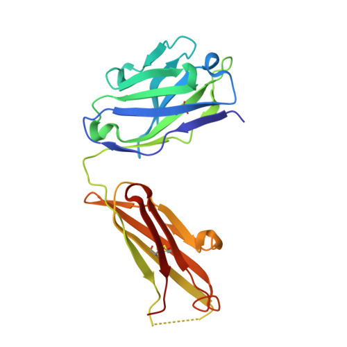
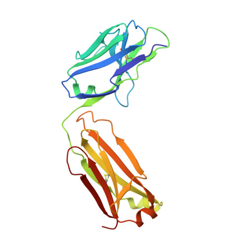
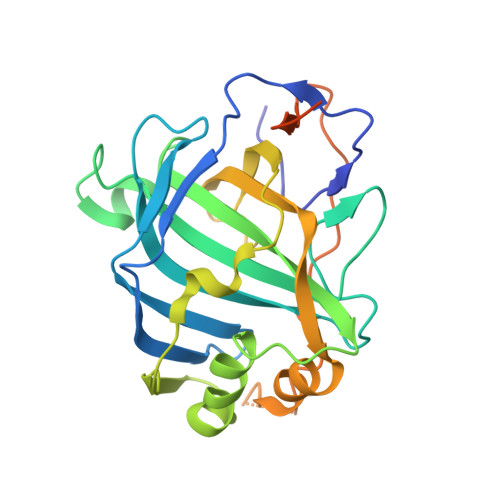
Structural and Biochemical Characterization of the Vaccinia Virus Envelope Protein D8 and Its Recognition by the Antibody LA5.

Matho, M.H.Maybeno, M.Benhnia, M.R.Becker, D.Meng, X.Xiang, Y.Crotty, S.Peters, B.Zajonc, D.M.

(2012) J Virol 86: 8050-8058

  • DOI: https://doi.org/10.1128/JVI.00836-12
  • Primary Citation Related Structures: 
    4E9O, 4EBQ, 4ETQ

  • PubMed Abstract: 

    Smallpox vaccine is considered a gold standard of vaccines, as it is the only one that has led to the complete eradication of an infectious disease from the human population. B cell responses are critical for the protective immunity induced by the vaccine, yet their targeted epitopes recognized in humans remain poorly described. Here we describe the biochemical and structural characterization of one of the immunodominant vaccinia virus (VACV) antigens, D8, and its binding to the monoclonal antibody LA5, which is capable of neutralizing VACV in the presence of complement. The full-length D8 ectodomain was found to form a tetramer. We determined the crystal structure of the LA5 Fab-monomeric D8 complex at a resolution of 2.1 Å, as well as the unliganded structures of D8 and LA5-Fab at resolutions of 1.42 Å and 1.6 Å, respectively. D8 features a carbonic anhydrase (CAH) fold that has evolved to bind to the glycosaminoglycan (GAG) chondroitin sulfate (CS) on host cells. The central positively charged crevice of D8 was predicted to be the CS binding site by automated docking experiments. Furthermore, sequence alignment of various poxvirus D8 orthologs revealed that this crevice is structurally conserved. The D8 epitope is formed by 23 discontinuous residues that are spread across 80% of the D8 protein sequence. Interestingly, LA5 binds with a high-affinity lock-and-key mechanism above this crevice with an unusually large antibody-antigen interface, burying 2,434 Å(2) of protein surface.


  • Organizational Affiliation
    • Division of Cell Biology, Division of Vaccine Discovery, La Jolla Institute for Allergy and Immunology, La Jolla, California, USA.

Macromolecule Content 

  • Total Structure Weight: 157.83 kDa 
  • Atom Count: 10,798 
  • Modeled Residue Count: 1,309 
  • Deposited Residue Count: 1,404 
  • Unique protein chains: 3

Macromolecules

Find similar proteins by:|  3D Structure
Entity ID: 1
MoleculeChains  Sequence LengthOrganismDetailsImage
anti-vaccinia D8L antigen murine monoclonal IgG2a antibody LA5, heavy chainA [auth H],
C [auth A]
221Mus musculusMutation(s): 0 
Gene Names: IgG2aIGHV1S127*01
Find similar proteins by:|  3D Structure
Entity ID: 2
MoleculeChains  Sequence LengthOrganismDetailsImage
anti-vaccinia D8L antigen murine monoclonal IgG2a antibody LA5, light chainB [auth L],
D [auth B]
212Mus musculusMutation(s): 0 
Gene Names: IgG2aIGKV4-55*01 germ line
Find similar proteins by:|  3D Structure
Entity ID: 3
MoleculeChains  Sequence LengthOrganismDetailsImage
IMV membrane proteinE [auth X],
F [auth C]
269Orthopoxvirus vacciniaMutation(s): 0 
Gene Names: D8LVACAC2_124VACCL3_124VAC_DPP17_124
UniProt
Find proteins for Q1M1K6 (Vaccinia virus)
Explore Q1M1K6 
Go to UniProtKB:  Q1M1K6
Entity Groups
Sequence Clusters30% Identity50% Identity70% Identity90% Identity95% Identity100% Identity
UniProt GroupQ1M1K6
Sequence Annotations
Expand
Reference Sequence

Small Molecules

Ligands 4 Unique
IDChains Name / Formula / InChI Key2D Diagram3D Interactions
GOL

Query on GOL


Download:Ideal Coordinates CCD File
I [auth L],
K [auth A],
N [auth B]
GLYCEROL
C3 H8 O3
PEDCQBHIVMGVHV-UHFFFAOYSA-N
EDO

Query on EDO


Download:Ideal Coordinates CCD File
H
1,2-ETHANEDIOL
C2 H6 O2
LYCAIKOWRPUZTN-UHFFFAOYSA-N
SCN

Query on SCN


Download:Ideal Coordinates CCD File
G [auth H]
J [auth L]
L [auth A]
M [auth A]
O [auth B]
G [auth H],
J [auth L],
L [auth A],
M [auth A],
O [auth B],
P [auth X]
THIOCYANATE ION
C N S
ZMZDMBWJUHKJPS-UHFFFAOYSA-M
CL

Query on CL


Download:Ideal Coordinates CCD File
Q [auth C]CHLORIDE ION
Cl
VEXZGXHMUGYJMC-UHFFFAOYSA-M

Experimental Data & Validation

Experimental Data

  • Method: X-RAY DIFFRACTION
  • Resolution: 2.10 Å
  • R-Value Free:  0.252 (Depositor), 0.253 (DCC) 
  • R-Value Work:  0.202 (Depositor), 0.202 (DCC) 
  • R-Value Observed: 0.203 (Depositor) 
Space Group: P 1 21 1
Unit Cell:
Length ( Å )Angle ( ˚ )
a = 76.021α = 90
b = 91.156β = 107.32
c = 103.774γ = 90
Software Package:
Software NamePurpose
DENZOdata reduction
SCALEPACKdata scaling
MOLREPphasing
REFMACrefinement
PDB_EXTRACTdata extraction

Structure Validation

View Full Validation Report


Entry History 

Deposition Data

Revision History  (Full details and data files)

  • Version 1.0: 2012-06-06
    Type: Initial release
  • Version 1.1: 2012-07-25
    Changes: Database references
  • Version 1.2: 2023-09-13
    Changes: Data collection, Database references, Derived calculations, Refinement description
  • Version 1.3: 2024-11-20
    Changes: Structure summary