4ER6 | pdb_00004er6

Crystal structure of human DOT1L in complex with inhibitor SGC0946


Experimental Data Snapshot

  • Method: X-RAY DIFFRACTION
  • Resolution: 2.30 Å
  • R-Value Free: 
    0.230 (Depositor), 0.232 (DCC) 
  • R-Value Work: 
    0.216 (Depositor), 0.217 (DCC) 
  • R-Value Observed: 
    0.217 (Depositor) 

wwPDB Validation 3D Report Full Report

Validation slider image for 4ER6

Ligand Structure Quality Assessment 


This is version 1.3 of the entry. See complete history

Literature

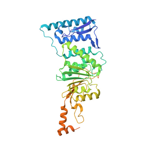
Catalytic site remodelling of the DOT1L methyltransferase by selective inhibitors.

Yu, W.Chory, E.J.Wernimont, A.K.Tempel, W.Scopton, A.Federation, A.Marineau, J.J.Qi, J.Barsyte-Lovejoy, D.Yi, J.Marcellus, R.Iacob, R.E.Engen, J.R.Griffin, C.Aman, A.Wienholds, E.Li, F.Pineda, J.Estiu, G.Shatseva, T.Hajian, T.Al-Awar, R.Dick, J.E.Vedadi, M.Brown, P.J.Arrowsmith, C.H.Bradner, J.E.Schapira, M.

(2012) Nat Commun 3: 1288-1288

  • DOI: https://doi.org/10.1038/ncomms2304
  • Primary Citation Related Structures: 
    3UWP, 4EQZ, 4ER0, 4ER3, 4ER5, 4ER6, 4ER7

  • PubMed Abstract: 

    Selective inhibition of protein methyltransferases is a promising new approach to drug discovery. An attractive strategy towards this goal is the development of compounds that selectively inhibit binding of the cofactor, S-adenosylmethionine, within specific protein methyltransferases. Here we report the three-dimensional structure of the protein methyltransferase DOT1L bound to EPZ004777, the first S-adenosylmethionine-competitive inhibitor of a protein methyltransferase with in vivo efficacy. This structure and those of four new analogues reveal remodelling of the catalytic site. EPZ004777 and a brominated analogue, SGC0946, inhibit DOT1L in vitro and selectively kill mixed lineage leukaemia cells, in which DOT1L is aberrantly localized via interaction with an oncogenic MLL fusion protein. These data provide important new insight into mechanisms of cell-active S-adenosylmethionine-competitive protein methyltransferase inhibitors, and establish a foundation for the further development of drug-like inhibitors of DOT1L for cancer therapy.


  • Organizational Affiliation
    • Structural Genomics Consortium, University of Toronto, Toronto, ON M5G 1L7, Canada.

Macromolecule Content 

  • Total Structure Weight: 47.77 kDa 
  • Atom Count: 2,682 
  • Modeled Residue Count: 335 
  • Deposited Residue Count: 413 
  • Unique protein chains: 1

Macromolecules

Find similar proteins by:|  3D Structure
Entity ID: 1
MoleculeChains  Sequence LengthOrganismDetailsImage
Histone-lysine N-methyltransferase, H3 lysine-79 specific413Homo sapiensMutation(s): 0 
Gene Names: DOT1LKIAA1814KMT4
EC: 2.1.1.43 (PDB Primary Data), 2.1.1.360 (UniProt)
UniProt & NIH Common Fund Data Resources
Find proteins for Q8TEK3 (Homo sapiens)
Explore Q8TEK3 
Go to UniProtKB:  Q8TEK3
PHAROS:  Q8TEK3
GTEx:  ENSG00000104885 
Entity Groups
Sequence Clusters30% Identity50% Identity70% Identity90% Identity95% Identity100% Identity
UniProt GroupQ8TEK3
Sequence Annotations
Expand
Reference Sequence

Small Molecules

Ligands 3 Unique
IDChains Name / Formula / InChI Key2D Diagram3D Interactions
AW2

Query on AW2


Download:Ideal Coordinates CCD File
C [auth A]5-bromo-7-{5-[(3-{[(4-tert-butylphenyl)carbamoyl]amino}propyl)(propan-2-yl)amino]-5-deoxy-beta-D-ribofuranosyl}-7H-pyrrolo[2,3-d]pyrimidin-4-amine
C28 H40 Br N7 O4
IQCKJUKAQJINMK-HUBRGWSESA-N
BR

Query on BR


Download:Ideal Coordinates CCD File
B [auth A]BROMIDE ION
Br
CPELXLSAUQHCOX-UHFFFAOYSA-M
NA

Query on NA


Download:Ideal Coordinates CCD File
D [auth A]SODIUM ION
Na
FKNQFGJONOIPTF-UHFFFAOYSA-N
Binding Affinity Annotations 
IDSourceBinding Affinity
AW2 BindingDB:  4ER6 Kd: min: 0.06, max: 5.6 (nM) from 2 assay(s)
IC50: min: 0.3, max: 8.8 (nM) from 2 assay(s)
EC50: 9 (nM) from 1 assay(s)

Experimental Data & Validation

Experimental Data

  • Method: X-RAY DIFFRACTION
  • Resolution: 2.30 Å
  • R-Value Free:  0.230 (Depositor), 0.232 (DCC) 
  • R-Value Work:  0.216 (Depositor), 0.217 (DCC) 
  • R-Value Observed: 0.217 (Depositor) 
Space Group: P 65
Unit Cell:
Length ( Å )Angle ( ˚ )
a = 149.907α = 90
b = 149.907β = 90
c = 52.839γ = 120
Software Package:
Software NamePurpose
MOSFLMdata reduction
SCALAdata scaling
REFMACrefinement
PDB_EXTRACTdata extraction
31IDdata collection

Structure Validation

View Full Validation Report


Ligand Structure Quality Assessment 


Entry History 

Revision History  (Full details and data files)

  • Version 1.0: 2012-05-16
    Type: Initial release
  • Version 1.1: 2017-11-15
    Changes: Refinement description
  • Version 1.2: 2018-05-16
    Changes: Data collection, Database references
  • Version 1.3: 2024-02-28
    Changes: Data collection, Database references, Derived calculations