4ENZ | pdb_00004enz

Structure of human ceruloplasmin at 2.6 A resolution


Experimental Data Snapshot

  • Method: X-RAY DIFFRACTION
  • Resolution: 2.60 Å
  • R-Value Free: 
    0.234 (Depositor), 0.229 (DCC) 
  • R-Value Work: 
    0.199 (Depositor), 0.200 (DCC) 
  • R-Value Observed: 
    0.201 (Depositor) 

Starting Model: experimental
View more details

wwPDB Validation 3D Report Full Report

Validation slider image for 4ENZ

Ligand Structure Quality Assessment 


This is version 1.5 of the entry. See complete history

Literature

Ceruloplasmin: macromolecular assemblies with iron-containing acute phase proteins.

Samygina, V.R.Sokolov, A.V.Bourenkov, G.Petoukhov, M.V.Pulina, M.O.Zakharova, E.T.Vasilyev, V.B.Bartunik, H.Svergun, D.I.

(2013) PLoS One 8: e67145-e67145

  • DOI: https://doi.org/10.1371/journal.pone.0067145
  • Primary Citation Related Structures: 
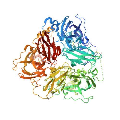
    4EJX, 4ENZ

  • PubMed Abstract: 

    Copper-containing ferroxidase ceruloplasmin (Cp) forms binary and ternary complexes with cationic proteins lactoferrin (Lf) and myeloperoxidase (Mpo) during inflammation. We present an X-ray crystal structure of a 2Cp-Mpo complex at 4.7 Å resolution. This structure allows one to identify major protein-protein interaction areas and provides an explanation for a competitive inhibition of Mpo by Cp and for the activation of p-phenylenediamine oxidation by Mpo. Small angle X-ray scattering was employed to construct low-resolution models of the Cp-Lf complex and, for the first time, of the ternary 2Cp-2Lf-Mpo complex in solution. The SAXS-based model of Cp-Lf supports the predicted 1:1 stoichiometry of the complex and demonstrates that both lobes of Lf contact domains 1 and 6 of Cp. The 2Cp-2Lf-Mpo SAXS model reveals the absence of interaction between Mpo and Lf in the ternary complex, so Cp can serve as a mediator of protein interactions in complex architecture. Mpo protects antioxidant properties of Cp by isolating its sensitive loop from proteases. The latter is important for incorporation of Fe(3+) into Lf, which activates ferroxidase activity of Cp and precludes oxidation of Cp substrates. Our models provide the structural basis for possible regulatory role of these complexes in preventing iron-induced oxidative damage.


  • Organizational Affiliation
    • Institute of Crystallography RAS, Moscow, Russia. vsamygina@cicbiogune.es

Macromolecule Content 

  • Total Structure Weight: 123.38 kDa 
  • Atom Count: 8,468 
  • Modeled Residue Count: 1,029 
  • Deposited Residue Count: 1,065 
  • Unique protein chains: 1

Macromolecules

Find similar proteins by:|  3D Structure
Entity ID: 1
MoleculeChains  Sequence LengthOrganismDetailsImage
Ceruloplasmin1,065Homo sapiensMutation(s): 0 
EC: 1.16.3.1 (PDB Primary Data), 1.16.3.4 (UniProt), 1.11.1.27 (UniProt), 1.11.1.9 (UniProt)
UniProt & NIH Common Fund Data Resources
Find proteins for P00450 (Homo sapiens)
Explore P00450 
Go to UniProtKB:  P00450
PHAROS:  P00450
GTEx:  ENSG00000047457 
Entity Groups
Sequence Clusters30% Identity50% Identity70% Identity90% Identity95% Identity100% Identity
UniProt GroupP00450
Glycosylation
Glycosylation Sites: 2Go to GlyGen: P00450-1
Sequence Annotations
Expand
Reference Sequence

Small Molecules

Ligands 7 Unique
IDChains Name / Formula / InChI Key2D Diagram3D Interactions
NAG

Query on NAG


Download:Ideal Coordinates CCD File
B [auth A],
L [auth A]
2-acetamido-2-deoxy-beta-D-glucopyranose
C8 H15 N O6
OVRNDRQMDRJTHS-FMDGEEDCSA-N
GOL

Query on GOL


Download:Ideal Coordinates CCD File
N [auth A]GLYCEROL
C3 H8 O3
PEDCQBHIVMGVHV-UHFFFAOYSA-N
CU

Query on CU


Download:Ideal Coordinates CCD File
C [auth A]
D [auth A]
E [auth A]
F [auth A]
G [auth A]
C [auth A],
D [auth A],
E [auth A],
F [auth A],
G [auth A],
H [auth A]
COPPER (II) ION
Cu
JPVYNHNXODAKFH-UHFFFAOYSA-N
CA

Query on CA


Download:Ideal Coordinates CCD File
K [auth A]CALCIUM ION
Ca
BHPQYMZQTOCNFJ-UHFFFAOYSA-N
OXY

Query on OXY


Download:Ideal Coordinates CCD File
J [auth A]OXYGEN MOLECULE
O2
MYMOFIZGZYHOMD-UHFFFAOYSA-N
NA

Query on NA


Download:Ideal Coordinates CCD File
M [auth A]SODIUM ION
Na
FKNQFGJONOIPTF-UHFFFAOYSA-N
O

Query on O


Download:Ideal Coordinates CCD File
I [auth A]OXYGEN ATOM
O
XLYOFNOQVPJJNP-UHFFFAOYSA-N

Experimental Data & Validation

Experimental Data

  • Method: X-RAY DIFFRACTION
  • Resolution: 2.60 Å
  • R-Value Free:  0.234 (Depositor), 0.229 (DCC) 
  • R-Value Work:  0.199 (Depositor), 0.200 (DCC) 
  • R-Value Observed: 0.201 (Depositor) 
Space Group: P 32 2 1
Unit Cell:
Length ( Å )Angle ( ˚ )
a = 210.775α = 90
b = 210.775β = 90
c = 84.502γ = 120
Software Package:
Software NamePurpose
PROTEUM PLUSdata collection
MOLREPphasing
REFMACrefinement
DENZOdata reduction
SCALEPACKdata scaling

Structure Validation

View Full Validation Report


Ligand Structure Quality Assessment 


Entry History 

Deposition Data

Revision History  (Full details and data files)

  • Version 1.0: 2013-04-17
    Type: Initial release
  • Version 1.1: 2013-07-17
    Changes: Database references
  • Version 1.2: 2013-09-11
    Changes: Database references
  • Version 1.3: 2020-07-29
    Type: Remediation
    Reason: Carbohydrate remediation
    Changes: Data collection, Derived calculations, Structure summary
  • Version 1.4: 2023-09-13
    Changes: Data collection, Database references, Refinement description, Structure summary
  • Version 1.5: 2024-11-27
    Changes: Structure summary