4EMV | pdb_00004emv

Crystal structure of a topoisomerase ATP inhibitor


Experimental Data Snapshot

  • Method: X-RAY DIFFRACTION
  • Resolution: 1.70 Å
  • R-Value Free: 
    0.223 (Depositor), 0.226 (DCC) 
  • R-Value Work: 
    0.189 (Depositor), 0.196 (DCC) 
  • R-Value Observed: 
    0.191 (Depositor) 

wwPDB Validation 3D Report Full Report

Validation slider image for 4EMV

Ligand Structure Quality Assessment 


This is version 1.1 of the entry. See complete history

Literature

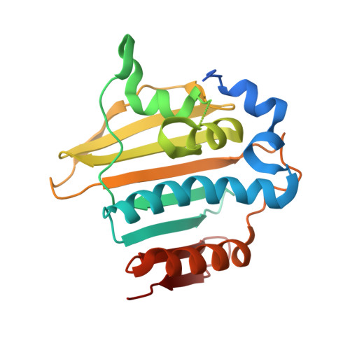
Discovery of a novel azaindole class of antibacterial agents targeting the ATPase domains of DNA gyrase and Topoisomerase IV.

Manchester, J.I.Dussault, D.D.Rose, J.A.Boriack-Sjodin, P.A.Uria-Nickelsen, M.Ioannidis, G.Bist, S.Fleming, P.Hull, K.G.

(2012) Bioorg Med Chem Lett 22: 5150-5156

  • DOI: https://doi.org/10.1016/j.bmcl.2012.05.128
  • Primary Citation Related Structures: 
    4EM7, 4EMV

  • PubMed Abstract: 

    We present the discovery and optimization of a novel series of bacterial topoisomerase inhibitors. Starting from a virtual screening hit, activity was optimized through a combination of structure-based design and physical property optimization. Synthesis of fewer than a dozen compounds was required to achieve inhibition of the growth of methicillin-resistant Staphyloccus aureus (MRSA) at compound concentrations of 1.56 μM. These compounds simultaneously inhibit DNA gyrase and Topoisomerase IV at similar nanomolar concentrations, reducing the likelihood of the spontaneous occurrence of target-based mutations resulting in antibiotic resistance, an increasing threat in the treatment of serious infections.


  • Organizational Affiliation
    • Infection Innovative Medicines Unit, AstraZeneca R&D Boston, Waltham, MA 02451, USA. john.manchester@astrazeneca.com

Macromolecule Content 

  • Total Structure Weight: 25.05 kDa 
  • Atom Count: 1,760 
  • Modeled Residue Count: 196 
  • Deposited Residue Count: 226 
  • Unique protein chains: 1

Macromolecules

Find similar proteins by:|  3D Structure
Entity ID: 1
MoleculeChains  Sequence LengthOrganismDetailsImage
DNA topoisomerase IV, B subunit226Streptococcus pneumoniae GA47373Mutation(s): 0 
Gene Names: ParESPAR94_0831
EC: 5.99.1

Small Molecules

Ligands 1 Unique
IDChains Name / Formula / InChI Key2D Diagram3D Interactions
0R9

Query on 0R9


Download:Ideal Coordinates CCD File
B [auth A]5-{2-(ethylcarbamoyl)-4-[3-(trifluoromethyl)-1H-pyrazol-1-yl]-1H-pyrrolo[2,3-b]pyridin-5-yl}pyridine-3-carboxylic acid
C20 H15 F3 N6 O3
QEODWPGJXCSBCR-UHFFFAOYSA-N

Experimental Data & Validation

Experimental Data

  • Method: X-RAY DIFFRACTION
  • Resolution: 1.70 Å
  • R-Value Free:  0.223 (Depositor), 0.226 (DCC) 
  • R-Value Work:  0.189 (Depositor), 0.196 (DCC) 
  • R-Value Observed: 0.191 (Depositor) 
Space Group: C 2 2 21
Unit Cell:
Length ( Å )Angle ( ˚ )
a = 73.297α = 90
b = 94.92β = 90
c = 61.247γ = 90
Software Package:
Software NamePurpose
MOSFLMdata reduction
SCALAdata scaling
AMoREphasing
REFMACrefinement
PDB_EXTRACTdata extraction

Structure Validation

View Full Validation Report


Ligand Structure Quality Assessment 


Entry History 

Deposition Data

Revision History  (Full details and data files)

  • Version 1.0: 2012-08-01
    Type: Initial release
  • Version 1.1: 2024-02-28
    Changes: Data collection, Database references, Derived calculations