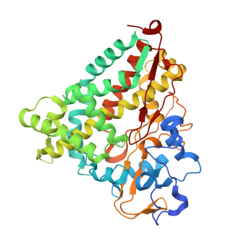
4EK1

Crystal Structure of Electron-Spin Labeled Cytochrome P450cam


Experimental Data Snapshot

  • Method: X-RAY DIFFRACTION
  • Resolution: 1.97 Å
  • R-Value Free: 0.253 
  • R-Value Work: 0.204 
  • R-Value Observed: 0.207 

wwPDB Validation   3D Report Full Report


Ligand Structure Quality Assessment 


This is version 1.2 of the entry. See complete history


Literature

Double electron-electron resonance shows cytochrome P450cam undergoes a conformational change in solution upon binding substrate.

Stoll, S.Lee, Y.T.Zhang, M.Wilson, R.F.Britt, R.D.Goodin, D.B.

(2012) Proc Natl Acad Sci U S A 109: 12888-12893

  • DOI: https://doi.org/10.1073/pnas.1207123109
  • Primary Citation of Related Structures:  
    4EK1

  • PubMed Abstract: 

    Although cytochrome P450cam from Pseudomonas putida, the archetype for all heme monooxygenases, has long been known to have a closed active site, recent reports show that the enzyme can also be crystallized in at least two clusters of open conformations. This suggests that the enzyme may undergo significant conformational changes during substrate binding and catalytic turnover. However, these conformations were observed in the crystalline state, and information is needed about the conformations that are populated in solution. In this study, double electron-electron resonance experiments were performed to observe substrate-induced changes in distance as measured by the dipolar coupling between spin labels introduced onto the surface of the enzyme on opposite sides of the substrate access channel. The double electron-electron resonance data show a decrease of 0.8 nm in the distance between spin labels placed at S48C and S190C upon binding the substrate camphor. A rotamer distribution model based on the crystal structures adequately describes the observed distance distributions. These results demonstrate conclusively that, in the physiologically relevant solution state, the substrate-free enzyme exists in the open P450cam-O conformation and that camphor binding results in conversion to the closed P450cam-C form. This approach should be useful for investigating many other P450s, including mammalian forms, in which the role of conformational change is of central importance but not well understood.


  • Organizational Affiliation

    Department of Chemistry, University of California, One Shields Avenue, Davis, CA 95616, USA.


Macromolecules
Find similar proteins by:  (by identity cutoff)  |  3D Structure
Entity ID: 1
MoleculeChains Sequence LengthOrganismDetailsImage
Camphor 5-monooxygenase
A, B
414Pseudomonas putidaMutation(s): 7 
Gene Names: camCcyp101
EC: 1.14.15.1
UniProt
Find proteins for P00183 (Pseudomonas putida)
Explore P00183 
Go to UniProtKB:  P00183
Entity Groups  
Sequence Clusters30% Identity50% Identity70% Identity90% Identity95% Identity100% Identity
UniProt GroupP00183
Sequence Annotations
Expand
  • Reference Sequence
Small Molecules
Ligands 4 Unique
IDChains Name / Formula / InChI Key2D Diagram3D Interactions
HEM
Query on HEM

Download Ideal Coordinates CCD File 
C [auth A],
H [auth B]
PROTOPORPHYRIN IX CONTAINING FE
C34 H32 Fe N4 O4
KABFMIBPWCXCRK-RGGAHWMASA-L
MTN
Query on MTN

Download Ideal Coordinates CCD File 
F [auth A],
G [auth A],
K [auth B]
S-[(1-oxyl-2,2,5,5-tetramethyl-2,5-dihydro-1H-pyrrol-3-yl)methyl] methanesulfonothioate
C10 H18 N O3 S2
MXZPGYFBZHBAQM-UHFFFAOYSA-N
CAM
Query on CAM

Download Ideal Coordinates CCD File 
D [auth A],
I [auth B]
CAMPHOR
C10 H16 O
DSSYKIVIOFKYAU-XCBNKYQSSA-N
K
Query on K

Download Ideal Coordinates CCD File 
E [auth A],
J [auth B]
POTASSIUM ION
K
NPYPAHLBTDXSSS-UHFFFAOYSA-N
Experimental Data & Validation

Experimental Data

  • Method: X-RAY DIFFRACTION
  • Resolution: 1.97 Å
  • R-Value Free: 0.253 
  • R-Value Work: 0.204 
  • R-Value Observed: 0.207 
  • Space Group: P 1 21 1
Unit Cell:
Length ( Å )Angle ( ˚ )
a = 56.02α = 90
b = 101.53β = 107.39
c = 72.98γ = 90
Software Package:
Software NamePurpose
MOSFLMdata reduction
SCALAdata scaling
MOLREPphasing
REFMACrefinement
PDB_EXTRACTdata extraction
Blu-Icedata collection

Structure Validation

View Full Validation Report


Ligand Structure Quality Assessment 


Entry History 

Deposition Data

Revision History  (Full details and data files)

  • Version 1.0: 2012-07-25
    Type: Initial release
  • Version 1.1: 2012-08-29
    Changes: Database references
  • Version 1.2: 2024-10-16
    Changes: Advisory, Data collection, Database references, Derived calculations, Structure summary