4EI5 | pdb_00004ei5

Crystal Structure of XV19 TCR in complex with CD1d-sulfatide C24:1


Experimental Data Snapshot

  • Method: X-RAY DIFFRACTION
  • Resolution: 3.10 Å
  • R-Value Free: 
    0.227 (Depositor), 0.248 (DCC) 
  • R-Value Work: 
    0.197 (Depositor), 0.213 (DCC) 
  • R-Value Observed: 
    0.199 (Depositor) 

Starting Models: experimental
View more details

wwPDB Validation 3D Report Full Report

Validation slider image for 4EI5

Ligand Structure Quality Assessment 


This is version 2.2 of the entry. See complete history

Literature

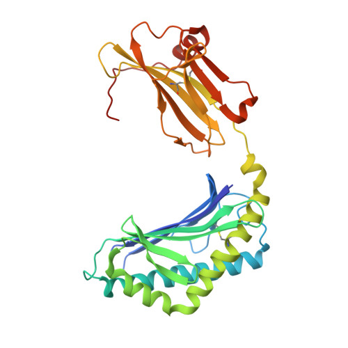
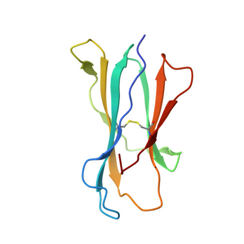
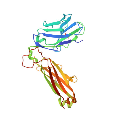
Recognition of CD1d-sulfatide mediated by a type II natural killer T cell antigen receptor.

Patel, O.Pellicci, D.G.Gras, S.Sandoval-Romero, M.L.Uldrich, A.P.Mallevaey, T.Clarke, A.J.Le Nours, J.Theodossis, A.Cardell, S.L.Gapin, L.Godfrey, D.I.Rossjohn, J.

(2012) Nat Immunol 13: 857-863

  • DOI: https://doi.org/10.1038/ni.2372
  • Primary Citation Related Structures: 
    4EI5, 4EI6

  • PubMed Abstract: 

    Natural killer T cells (NKT cells) are divided into type I and type II subsets on the basis of differences in their T cell antigen receptor (TCR) repertoire and CD1d-antigen specificity. Although the mode by which type I NKT cell TCRs recognize CD1d-antigen has been established, how type II NKT cell TCRs engage CD1d-antigen is unknown. Here we provide a basis for how a type II NKT cell TCR, XV19, recognized CD1d-sulfatide. The XV19 TCR bound orthogonally above the A' pocket of CD1d, in contrast to the parallel docking of type I NKT cell TCRs over the F' pocket of CD1d. At the XV19 TCR-CD1d-sulfatide interface, the TCRα and TCRβ chains sat centrally on CD1d, where the malleable CDR3 loops dominated interactions with CD1d-sulfatide. Accordingly, we highlight the diverse mechanisms by which NKT cell TCRs can bind CD1d and account for the distinct antigen specificity of type II NKT cells.


  • Organizational Affiliation
    • 1] Department of Biochemistry and Molecular Biology, School of Biomedical Sciences, Monash University, Clayton, Australia.

Macromolecule Content 

  • Total Structure Weight: 199.82 kDa 
  • Atom Count: 13,206 
  • Modeled Residue Count: 1,613 
  • Deposited Residue Count: 1,708 
  • Unique protein chains: 4

Macromolecules

Find similar proteins by:|  3D Structure
Entity ID: 1
MoleculeChains  Sequence LengthOrganismDetailsImage
Antigen-presenting glycoprotein CD1d1
A, E
302Mus musculusMutation(s): 0 
Gene Names: Cd1d1Cd1.1
UniProt & NIH Common Fund Data Resources
Find proteins for P11609 (Mus musculus)
Explore P11609 
Go to UniProtKB:  P11609
IMPC:  MGI:107674
Entity Groups
Sequence Clusters30% Identity50% Identity70% Identity90% Identity95% Identity100% Identity
UniProt GroupP11609
Glycosylation
Glycosylation Sites: 3Go to GlyGen: P11609-1
Sequence Annotations
Expand
Reference Sequence
Find similar proteins by:|  3D Structure
Entity ID: 2
MoleculeChains  Sequence LengthOrganismDetailsImage
Beta-2-microglobulin
B, F
99Mus musculusMutation(s): 0 
Gene Names: B2m
UniProt & NIH Common Fund Data Resources
Find proteins for P01887 (Mus musculus)
Explore P01887 
Go to UniProtKB:  P01887
IMPC:  MGI:88127
Entity Groups
Sequence Clusters30% Identity50% Identity70% Identity90% Identity95% Identity100% Identity
UniProt GroupP01887
Sequence Annotations
Expand
Reference Sequence
Find similar proteins by:|  3D Structure
Entity ID: 3
MoleculeChains  Sequence LengthOrganismDetailsImage
Valpha1 XV19 Type II Natural Killer T cell receptor (mouse variable domain, human constant domain)
C, G
208Mus musculusHomo sapiens
This entity is chimeric
Mutation(s): 0 
Find similar proteins by:|  3D Structure
Entity ID: 4
MoleculeChains  Sequence LengthOrganismDetailsImage
Vbeta16 XV19 Type II Natural Killer T cell receptor (mouse variable domain, human constant domain)
D, H
245Mus musculusHomo sapiens
This entity is chimeric
Mutation(s): 0 

Oligosaccharides

Help  
Entity ID: 5
MoleculeChains Length2D Diagram GlycosylationD Interactions
alpha-D-mannopyranose-(1-3)-beta-D-mannopyranose-(1-4)-2-acetamido-2-deoxy-beta-D-glucopyranose-(1-4)-2-acetamido-2-deoxy-beta-D-glucopyranose
I
4N-Glycosylation
Glycosylation Resources
GlyTouCan: G81315DD
GlyCosmos: G81315DD
GlyGen: G81315DD
Entity ID: 6
MoleculeChains Length2D Diagram GlycosylationD Interactions
2-acetamido-2-deoxy-beta-D-glucopyranose-(1-4)-2-acetamido-2-deoxy-beta-D-glucopyranose
J, L
2N-Glycosylation
Glycosylation Resources
GlyTouCan: G42666HT
GlyCosmos: G42666HT
GlyGen: G42666HT
Entity ID: 7
MoleculeChains Length2D Diagram GlycosylationD Interactions
beta-D-mannopyranose-(1-4)-2-acetamido-2-deoxy-beta-D-glucopyranose-(1-4)-2-acetamido-2-deoxy-beta-D-glucopyranose
K
3N-Glycosylation
Glycosylation Resources
GlyTouCan: G15407YE
GlyCosmos: G15407YE
GlyGen: G15407YE

Experimental Data & Validation

Experimental Data

  • Method: X-RAY DIFFRACTION
  • Resolution: 3.10 Å
  • R-Value Free:  0.227 (Depositor), 0.248 (DCC) 
  • R-Value Work:  0.197 (Depositor), 0.213 (DCC) 
  • R-Value Observed: 0.199 (Depositor) 
Space Group: P 21 21 21
Unit Cell:
Length ( Å )Angle ( ˚ )
a = 102.739α = 90
b = 140.711β = 90
c = 160.773γ = 90
Software Package:
Software NamePurpose
SCALAdata scaling
PHASERphasing
BUSTER-TNTrefinement
PDB_EXTRACTdata extraction
Blu-Icedata collection
XDSdata reduction
BUSTERrefinement

Structure Validation

View Full Validation Report


Ligand Structure Quality Assessment 


Entry History 

Deposition Data

Revision History  (Full details and data files)

  • Version 1.0: 2012-07-25
    Type: Initial release
  • Version 1.1: 2012-08-08
    Changes: Database references
  • Version 1.2: 2012-09-05
    Changes: Database references
  • Version 2.0: 2020-07-29
    Type: Remediation
    Reason: Carbohydrate remediation
    Changes: Advisory, Atomic model, Data collection, Database references, Derived calculations, Structure summary
  • Version 2.1: 2023-09-13
    Changes: Data collection, Database references, Refinement description, Structure summary
  • Version 2.2: 2024-11-27
    Changes: Structure summary