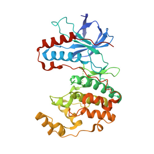
4EH9

Human p38 MAP kinase in complex with NP-F11 and RL87


Experimental Data Snapshot

  • Method: X-RAY DIFFRACTION
  • Resolution: 2.10 Å
  • R-Value Free: 0.280 
  • R-Value Work: 0.229 
  • R-Value Observed: 0.232 

Starting Model: experimental
View more details

wwPDB Validation   3D Report Full Report


Ligand Structure Quality Assessment 


This is version 1.2 of the entry. See complete history


Literature

Natural-product-derived fragments for fragment-based ligand discovery.

Over, B.Wetzel, S.Grutter, C.Nakai, Y.Renner, S.Rauh, D.Waldmann, H.

(2012) Nat Chem 5: 21-28

  • DOI: https://doi.org/10.1038/nchem.1506
  • Primary Citation of Related Structures:  
    4EH2, 4EH3, 4EH4, 4EH5, 4EH6, 4EH7, 4EH8, 4EH9, 4EHV

  • PubMed Abstract: 

    Fragment-based ligand and drug discovery predominantly employs sp(2)-rich compounds covering well-explored regions of chemical space. Despite the ease with which such fragments can be coupled, this focus on flat compounds is widely cited as contributing to the attrition rate of the drug discovery process. In contrast, biologically validated natural products are rich in stereogenic centres and populate areas of chemical space not occupied by average synthetic molecules. Here, we have analysed more than 180,000 natural product structures to arrive at 2,000 clusters of natural-product-derived fragments with high structural diversity, which resemble natural scaffolds and are rich in sp(3)-configured centres. The structures of the cluster centres differ from previously explored fragment libraries, but for nearly half of the clusters representative members are commercially available. We validate their usefulness for the discovery of novel ligand and inhibitor types by means of protein X-ray crystallography and the identification of novel stabilizers of inactive conformations of p38α MAP kinase and of inhibitors of several phosphatases.


  • Organizational Affiliation

    Max-Planck-Institute of Molecular Physiology, Otto-Hahn-Strasse 11, D-44227 Dortmund, Germany.


Macromolecules
Find similar proteins by:  (by identity cutoff)  |  3D Structure
Entity ID: 1
MoleculeChains Sequence LengthOrganismDetailsImage
Mitogen-activated protein kinase 14360Homo sapiensMutation(s): 0 
Gene Names: MAPK14CSBPCSBP1CSBP2CSPB1MXI2SAPK2A
EC: 2.7.11.24
UniProt & NIH Common Fund Data Resources
Find proteins for Q16539 (Homo sapiens)
Explore Q16539 
Go to UniProtKB:  Q16539
PHAROS:  Q16539
GTEx:  ENSG00000112062 
Entity Groups  
Sequence Clusters30% Identity50% Identity70% Identity90% Identity95% Identity100% Identity
UniProt GroupQ16539
Sequence Annotations
Expand
  • Reference Sequence
Small Molecules
Ligands 2 Unique
IDChains Name / Formula / InChI Key2D Diagram3D Interactions
0OQ
Query on 0OQ

Download Ideal Coordinates CCD File 
C [auth A](1R,5S)-3-[4-(trifluoromethyl)benzoyl]-1,2,3,4,5,6-hexahydro-8H-1,5-methanopyrido[1,2-a][1,5]diazocin-8-one
C19 H17 F3 N2 O2
FENJYCPCZAMGTO-GXTWGEPZSA-N
IRG
Query on IRG

Download Ideal Coordinates CCD File 
B [auth A]N~4~-cyclopropyl-2-phenylquinazoline-4,7-diamine
C17 H16 N4
WCANXWUISJCJOB-UHFFFAOYSA-N
Experimental Data & Validation

Experimental Data

  • Method: X-RAY DIFFRACTION
  • Resolution: 2.10 Å
  • R-Value Free: 0.280 
  • R-Value Work: 0.229 
  • R-Value Observed: 0.232 
  • Space Group: P 21 21 21
Unit Cell:
Length ( Å )Angle ( ˚ )
a = 65.62α = 90
b = 74.92β = 90
c = 78.19γ = 90
Software Package:
Software NamePurpose
XSCALEdata scaling
REFMACrefinement
PDB_EXTRACTdata extraction
XDSdata scaling
PHASERphasing

Structure Validation

View Full Validation Report


Ligand Structure Quality Assessment 


Entry History 

Deposition Data

Revision History  (Full details and data files)

  • Version 1.0: 2012-12-05
    Type: Initial release
  • Version 1.1: 2013-01-02
    Changes: Database references
  • Version 1.2: 2023-09-13
    Changes: Data collection, Database references, Derived calculations, Refinement description