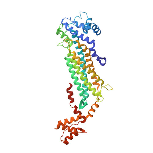
Crystal Structure of Adenylosuccinate Lyase from Francisella tularensis Complexed with AMP and Succinate
Maltseva, N., Kim, Y., Shatsman, S., Anderson, W.F., Joachimiak, A., Center for Structural Genomics of Infectious Diseases (CSGID)To be published.