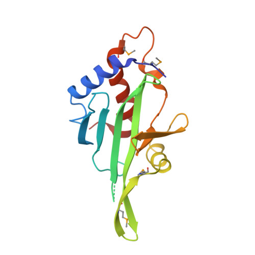
4EBR

Crystal structure of Autophagic E2, Atg10


Experimental Data Snapshot

  • Method: X-RAY DIFFRACTION
  • Resolution: 2.70 Å
  • R-Value Free: 0.251 
  • R-Value Work: 0.211 
  • R-Value Observed: 0.224 

wwPDB Validation   3D Report Full Report


This is version 1.2 of the entry. See complete history


Literature

Structure of the autophagic E2 enzyme Atg10

Hong, S.B.Kim, B.W.Kim, J.H.Song, H.K.

(2012) Acta Crystallogr D Biol Crystallogr 68: 1409-1417

  • DOI: https://doi.org/10.1107/S0907444912034166
  • Primary Citation of Related Structures:  
    4EBR

  • PubMed Abstract: 

    Autophagy is a regulated degradation pathway that plays a critical role in all eukaryotic life cycles. One interesting feature of the core autophagic process, autophagosome formation, is similar to ubiquitination. One of two autophagic E2 enzymes, Atg10, interacts with Atg7 to receive Atg12, a ubiquitin-like molecule, and is also involved in the Atg12-Atg5 conjugation reaction. To date, no information on the interaction between Atg10 and Atg7 has been reported, although structural information is available pertaining to the individual components. Here, the crystal structure of Atg10 from Saccharomyces cerevisiae is described at 2.7 Å resolution. A significant improvement of the diffraction limit by heavy-atom derivatization was essential for structure determination. The core fold of yeast Atg10 is well conserved compared with those of Atg3 and other E2 enzymes. In contrast to other E2 enzymes, however, the autophagic E2 enzymes Atg3 and Atg10 possess insertion regions in the middle of the core fold and may be involved in protein function. The missing segment, which was termed the `FR-region', in Atg10 may be important for interaction with the E1 enzyme Atg7. This study provides a framework for understanding the E2 conjugation reaction in autophagy.


  • Organizational Affiliation

    School of Life Sciences and Biotechnology, Korea University, Seoul 136-701, Republic of Korea.


Macromolecules
Find similar proteins by:  (by identity cutoff)  |  3D Structure
Entity ID: 1
MoleculeChains Sequence LengthOrganismDetailsImage
Ubiquitin-like-conjugating enzyme ATG10
A, B
169Saccharomyces cerevisiae S288CMutation(s): 0 
Gene Names: ATG10APG10YLL042C
EC: 6.3.2 (PDB Primary Data), 2.3.2 (UniProt)
UniProt
Find proteins for Q07879 (Saccharomyces cerevisiae (strain ATCC 204508 / S288c))
Explore Q07879 
Go to UniProtKB:  Q07879
Entity Groups  
Sequence Clusters30% Identity50% Identity70% Identity90% Identity95% Identity100% Identity
UniProt GroupQ07879
Sequence Annotations
Expand
  • Reference Sequence
Small Molecules
Modified Residues  1 Unique
IDChains TypeFormula2D DiagramParent
MSE
Query on MSE
A, B
L-PEPTIDE LINKINGC5 H11 N O2 SeMET
Experimental Data & Validation

Experimental Data

  • Method: X-RAY DIFFRACTION
  • Resolution: 2.70 Å
  • R-Value Free: 0.251 
  • R-Value Work: 0.211 
  • R-Value Observed: 0.224 
  • Space Group: P 61 2 2
Unit Cell:
Length ( Å )Angle ( ˚ )
a = 127.833α = 90
b = 127.833β = 90
c = 169.589γ = 120
Software Package:
Software NamePurpose
HKL-2000data collection
SOLVEphasing
PHENIXrefinement
HKL-2000data reduction
HKL-2000data scaling

Structure Validation

View Full Validation Report


Entry History 

Deposition Data

Revision History  (Full details and data files)

  • Version 1.0: 2012-10-03
    Type: Initial release
  • Version 1.1: 2013-07-24
    Changes: Database references
  • Version 1.2: 2024-10-30
    Changes: Data collection, Database references, Derived calculations, Structure summary