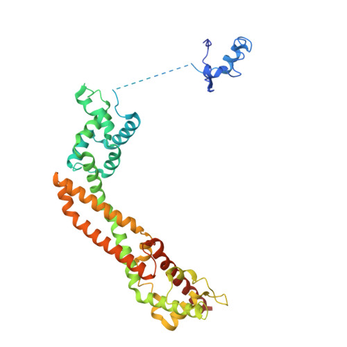
4EAH

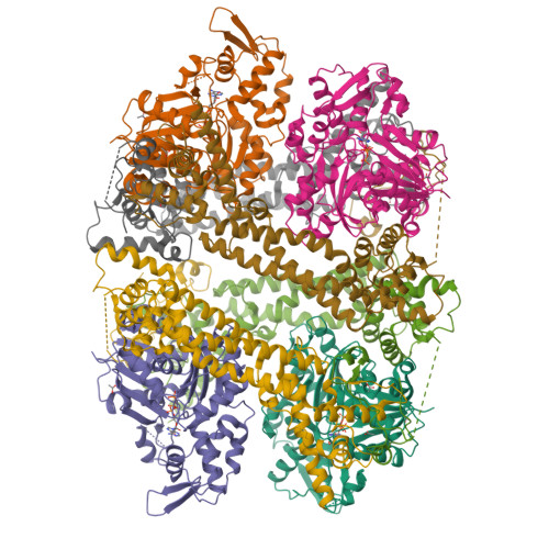
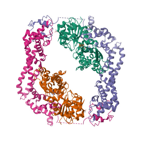
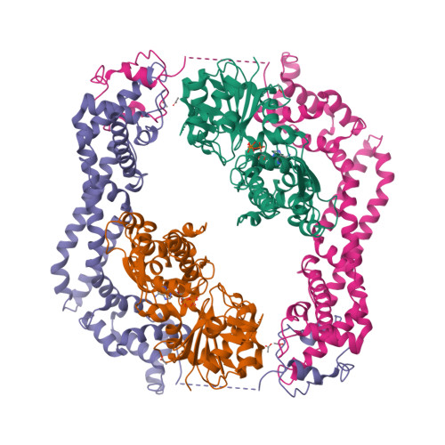
Crystal structure of the formin homology 2 domain of FMNL3 bound to actin


Experimental Data Snapshot

  • Method: X-RAY DIFFRACTION
  • Resolution: 3.40 Å
  • R-Value Free: 0.277 
  • R-Value Work: 0.230 
  • R-Value Observed: 0.233 

wwPDB Validation   3D Report Full Report


Ligand Structure Quality Assessment 


This is version 1.4 of the entry. See complete history


Literature

FMNL3 FH2-actin structure gives insight into formin-mediated actin nucleation and elongation.

Thompson, M.E.Heimsath, E.G.Gauvin, T.J.Higgs, H.N.Kull, F.J.

(2013) Nat Struct Mol Biol 20: 111-118

  • DOI: https://doi.org/10.1038/nsmb.2462
  • Primary Citation of Related Structures:  
    4EAH

  • PubMed Abstract: 

    Formins are actin-assembly factors that act in a variety of actin-based processes. The conserved formin homology 2 (FH2) domain promotes filament nucleation and influences elongation through interaction with the barbed end. FMNL3 is a formin that induces assembly of filopodia but whose FH2 domain is a poor nucleator. The 3.4-Å structure of a mouse FMNL3 FH2 dimer in complex with tetramethylrhodamine-actin uncovers details of formin-regulated actin elongation. We observe distinct FH2 actin-binding regions; interactions in the knob and coiled-coil subdomains are necessary for actin binding, whereas those in the lasso-post interface are important for the stepping mechanism. Biochemical and cellular experiments test the importance of individual residues for function. This structure provides details for FH2-mediated filament elongation by processive capping and supports a model in which C-terminal non-FH2 residues of FMNL3 are required to stabilize the filament nucleus.


  • Organizational Affiliation

    Department of Biochemistry, Geisel School of Medicine at Dartmouth, Hanover, New Hampshire, USA.


Macromolecules
Find similar proteins by:  (by identity cutoff)  |  3D Structure
Entity ID: 1
MoleculeChains Sequence LengthOrganismDetailsImage
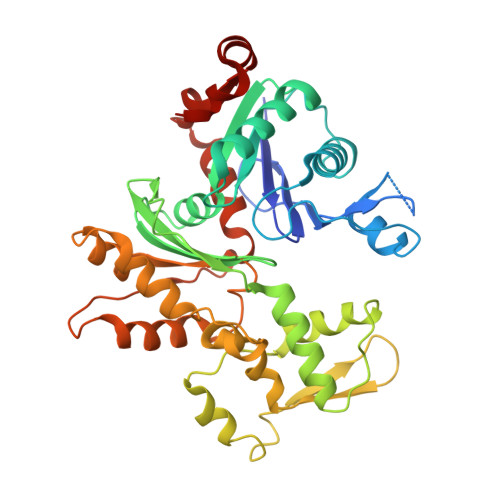
Actin, alpha skeletal muscleA [auth D],
F [auth H],
G,
H [auth F]
377Oryctolagus cuniculusMutation(s): 0 
EC: 3.6.4
UniProt
Find proteins for P68135 (Oryctolagus cuniculus)
Explore P68135 
Go to UniProtKB:  P68135
Entity Groups  
Sequence Clusters30% Identity50% Identity70% Identity90% Identity95% Identity100% Identity
UniProt GroupP68135
Sequence Annotations
Expand
  • Reference Sequence
Find similar proteins by:  (by identity cutoff)  |  3D Structure
Entity ID: 2
MoleculeChains Sequence LengthOrganismDetailsImage
Formin-like protein 3B [auth A],
C [auth E],
D [auth C],
E [auth B]
402Mus musculusMutation(s): 0 
Gene Names: Fmnl3Kiaa2014
UniProt & NIH Common Fund Data Resources
Find proteins for Q6ZPF4 (Mus musculus)
Explore Q6ZPF4 
Go to UniProtKB:  Q6ZPF4
IMPC:  MGI:109569
Entity Groups  
Sequence Clusters30% Identity50% Identity70% Identity90% Identity95% Identity100% Identity
UniProt GroupQ6ZPF4
Sequence Annotations
Expand
  • Reference Sequence
Small Molecules
Ligands 2 Unique
IDChains Name / Formula / InChI Key2D Diagram3D Interactions
ATP
Query on ATP

Download Ideal Coordinates CCD File 
J [auth D],
S [auth H],
U [auth G],
X [auth F]
ADENOSINE-5'-TRIPHOSPHATE
C10 H16 N5 O13 P3
ZKHQWZAMYRWXGA-KQYNXXCUSA-N
ACT
Query on ACT

Download Ideal Coordinates CCD File 
I [auth D]
K [auth A]
L [auth A]
M [auth A]
N [auth E]
I [auth D],
K [auth A],
L [auth A],
M [auth A],
N [auth E],
O [auth C],
P [auth C],
Q [auth B],
R [auth H],
T [auth G],
V [auth F],
W [auth F]
ACETATE ION
C2 H3 O2
QTBSBXVTEAMEQO-UHFFFAOYSA-M
Experimental Data & Validation

Experimental Data

  • Method: X-RAY DIFFRACTION
  • Resolution: 3.40 Å
  • R-Value Free: 0.277 
  • R-Value Work: 0.230 
  • R-Value Observed: 0.233 
  • Space Group: P 1 21 1
Unit Cell:
Length ( Å )Angle ( ˚ )
a = 125.98α = 90
b = 126.05β = 93.17
c = 129.62γ = 90
Software Package:
Software NamePurpose
JBluIce-EPICSdata collection
PHENIXmodel building
PHENIXrefinement
XDSdata reduction
XDSdata scaling
PHENIXphasing

Structure Validation

View Full Validation Report


Ligand Structure Quality Assessment 


Entry History 

Deposition Data

Revision History  (Full details and data files)

  • Version 1.0: 2012-12-12
    Type: Initial release
  • Version 1.1: 2012-12-26
    Changes: Database references
  • Version 1.2: 2013-01-16
    Changes: Database references
  • Version 1.3: 2017-11-15
    Changes: Advisory, Refinement description
  • Version 1.4: 2024-02-28
    Changes: Advisory, Data collection, Database references, Derived calculations