4E6U | pdb_00004e6u

Structure of LpxA from Acinetobacter baumannii at 1.4A resolution (P63 form)


Experimental Data Snapshot

  • Method: X-RAY DIFFRACTION
  • Resolution: 1.41 Å
  • R-Value Free: 
    0.191 (Depositor), 0.188 (DCC) 
  • R-Value Work: 
    0.175 (Depositor), 0.172 (DCC) 
  • R-Value Observed: 
    0.176 (Depositor) 

wwPDB Validation 3D Report Full Report

Validation slider image for 4E6U

This is version 1.1 of the entry. See complete history

Literature

Structure determination of LpxA from the lipopolysaccharide-synthesis pathway of Acinetobacter baumannii.

Badger, J.Chie-Leon, B.Logan, C.Sridhar, V.Sankaran, B.Zwart, P.H.Nienaber, V.

(2012) Acta Crystallogr Sect F Struct Biol Cryst Commun 68: 1477-1481

  • DOI: https://doi.org/10.1107/S174430911204571X
  • Primary Citation Related Structures: 
    4E6T, 4E6U

  • PubMed Abstract: 

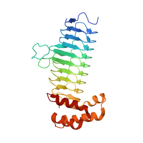
    Acinetobacter baumannii is a Gram-negative pathogenic bacterium which is resistant to most currently available antibiotics and that poses a significant health threat to hospital patients. LpxA is a key enzyme in the biosynthetic pathway of the lipopolysaccharides that are components of the bacterial outer membrane. It is a potential target for antibacterial agents that might be used to fight A. baumannii infections. This paper describes the structure determination of the apo form of LpxA in space groups P2(1)2(1)2(1) and P6(3). These crystal forms contained three and one protein molecules in the asymmetric unit and diffracted to 1.8 and 1.4 Å resolution, respectively. A comparison of the conformations of the independent protein monomers within and between the two crystal asymmetric units revealed very little structural variation across this set of structures. In the P6(3) crystal form the enzymatic site is exposed and is available for the introduction of small molecules of the type used in fragment-based drug discovery and structure-based lead optimization.


  • Organizational Affiliation
    • Zenobia Therapeutics Inc., 505 Coast Boulevard South, Suite 111, La Jolla, CA 92037, USA. john@zenobiatherapeutics.com

Macromolecule Content 

  • Total Structure Weight: 29.22 kDa 
  • Atom Count: 2,327 
  • Modeled Residue Count: 258 
  • Deposited Residue Count: 265 
  • Unique protein chains: 1

Macromolecules

Find similar proteins by:|  3D Structure
Entity ID: 1
MoleculeChains  Sequence LengthOrganismDetailsImage
Acyl-[acyl-carrier-protein]--UDP-N-acetylglucosamine O-acyltransferase265Acinetobacter baumanniiMutation(s): 0 
Gene Names: lpxAABTW07_2294
EC: 2.3.1.129

Small Molecules

Ligands 2 Unique
IDChains Name / Formula / InChI Key2D Diagram3D Interactions
SO4

Query on SO4


Download:Ideal Coordinates CCD File
B [auth A],
C [auth A]
SULFATE ION
O4 S
QAOWNCQODCNURD-UHFFFAOYSA-L
EDO

Query on EDO


Download:Ideal Coordinates CCD File
D [auth A]
E [auth A]
F [auth A]
G [auth A]
H [auth A]
D [auth A],
E [auth A],
F [auth A],
G [auth A],
H [auth A],
I [auth A],
J [auth A],
K [auth A],
L [auth A]
1,2-ETHANEDIOL
C2 H6 O2
LYCAIKOWRPUZTN-UHFFFAOYSA-N

Experimental Data & Validation

Experimental Data

  • Method: X-RAY DIFFRACTION
  • Resolution: 1.41 Å
  • R-Value Free:  0.191 (Depositor), 0.188 (DCC) 
  • R-Value Work:  0.175 (Depositor), 0.172 (DCC) 
  • R-Value Observed: 0.176 (Depositor) 
Space Group: P 63
Unit Cell:
Length ( Å )Angle ( ˚ )
a = 75.751α = 90
b = 75.751β = 90
c = 119.063γ = 120
Software Package:
Software NamePurpose
PHASERphasing
REFMACrefinement
HKL-2000data reduction
HKL-2000data scaling

Structure Validation

View Full Validation Report


Entry History 

Deposition Data

Revision History  (Full details and data files)

  • Version 1.0: 2012-12-12
    Type: Initial release
  • Version 1.1: 2024-02-28
    Changes: Data collection, Database references, Derived calculations