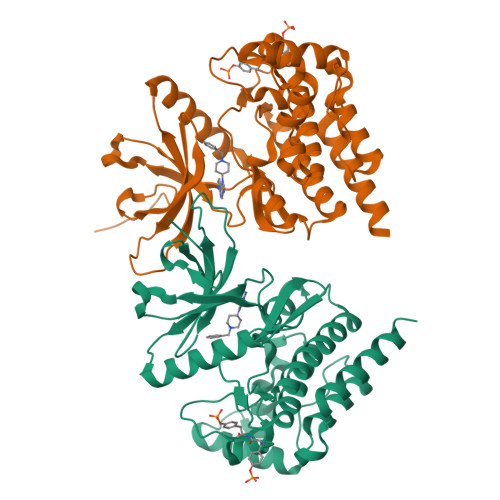
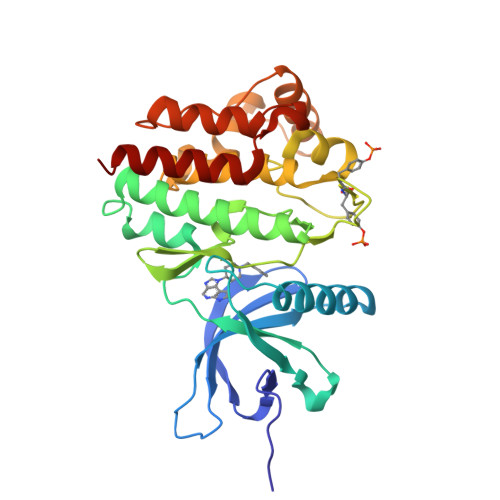
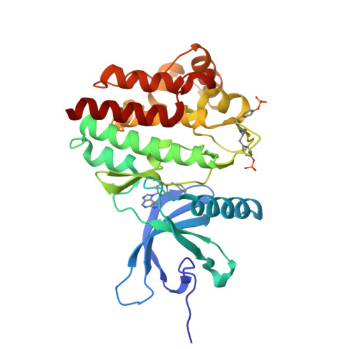
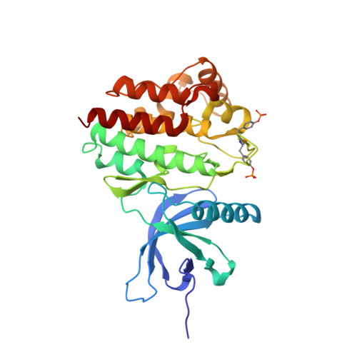
4E6Q

JAK2 kinase (JH1 domain) triple mutant in complex with compound 12


Experimental Data Snapshot

  • Method: X-RAY DIFFRACTION
  • Resolution: 1.95 Å
  • R-Value Free: 0.234 
  • R-Value Work: 0.186 
  • R-Value Observed: 0.189 

wwPDB Validation   3D Report Full Report


Ligand Structure Quality Assessment 


This is version 1.2 of the entry. See complete history


Literature

Identification of Imidazo-Pyrrolopyridines as Novel and Potent JAK1 Inhibitors.

Kulagowski, J.J.Blair, W.Bull, R.J.Chang, C.Deshmukh, G.Dyke, H.J.Eigenbrot, C.Ghilardi, N.Gibbons, P.Harrison, T.K.Hewitt, P.R.Liimatta, M.Hurley, C.A.Johnson, A.Johnson, T.Kenny, J.R.Bir Kohli, P.Maxey, R.J.Mendonca, R.Mortara, K.Murray, J.Narukulla, R.Shia, S.Steffek, M.Ubhayakar, S.Ultsch, M.van Abbema, A.Ward, S.I.Waszkowycz, B.Zak, M.

(2012) J Med Chem 55: 5901-5921

  • DOI: https://doi.org/10.1021/jm300438j
  • Primary Citation of Related Structures:  
    4E4L, 4E4M, 4E4N, 4E5W, 4E6D, 4E6Q

  • PubMed Abstract: 

    A therapeutic rationale is proposed for the treatment of inflammatory diseases, such as rheumatoid arthritis (RA), by specific targeting of the JAK1 pathway. Examination of the preferred binding conformation of clinically effective, pan-JAK inhibitor 1 led to identification of a novel, tricyclic hinge binding scaffold 3. Exploration of SAR through a series of cycloamino and cycloalkylamino analogues demonstrated this template to be highly tolerant of substitution, with a predisposition to moderate selectivity for the JAK1 isoform over JAK2. This study culminated in the identification of subnanomolar JAK1 inhibitors such as 22 and 49, having excellent cell potency, good rat pharmacokinetic characteristics, and excellent kinase selectivity. Determination of the binding modes of the series in JAK1 and JAK2 by X-ray crystallography supported the design of analogues to enhance affinity and selectivity.


  • Organizational Affiliation

    Department of Medicinal Chemistry, Argenta, 8/9 Spire Green Centre, Harlow CM19 5TR, United Kingdom. janusz.kulagowski@glpg


Macromolecules
Find similar proteins by:  (by identity cutoff)  |  3D Structure
Entity ID: 1
MoleculeChains Sequence LengthOrganismDetailsImage
Tyrosine-protein kinase JAK2
A, B
298Homo sapiensMutation(s): 3 
Gene Names: JAK2
EC: 2.7.10.2
UniProt & NIH Common Fund Data Resources
Find proteins for O60674 (Homo sapiens)
Explore O60674 
Go to UniProtKB:  O60674
PHAROS:  O60674
GTEx:  ENSG00000096968 
Entity Groups  
Sequence Clusters30% Identity50% Identity70% Identity90% Identity95% Identity100% Identity
UniProt GroupO60674
Sequence Annotations
Expand
  • Reference Sequence
Small Molecules
Ligands 1 Unique
IDChains Name / Formula / InChI Key2D Diagram3D Interactions
0NV
Query on 0NV

Download Ideal Coordinates CCD File 
C [auth A],
D [auth B]
1-(1-benzylpiperidin-4-yl)-1,6-dihydroimidazo[4,5-d]pyrrolo[2,3-b]pyridine
C20 H21 N5
WKWXJWGOAKGEEM-UHFFFAOYSA-N
Modified Residues  1 Unique
IDChains TypeFormula2D DiagramParent
PTR
Query on PTR
A, B
L-PEPTIDE LINKINGC9 H12 N O6 PTYR
Binding Affinity Annotations 
IDSourceBinding Affinity
0NV PDBBind:  4E6Q Ki: 4.4 (nM) from 1 assay(s)
Experimental Data & Validation

Experimental Data

  • Method: X-RAY DIFFRACTION
  • Resolution: 1.95 Å
  • R-Value Free: 0.234 
  • R-Value Work: 0.186 
  • R-Value Observed: 0.189 
  • Space Group: P 1 21 1
Unit Cell:
Length ( Å )Angle ( ˚ )
a = 50.502α = 90
b = 103.854β = 90.02
c = 69.999γ = 90
Software Package:
Software NamePurpose
d*TREKdata scaling
SCALAdata scaling
PHASERphasing
PHENIXrefinement
PDB_EXTRACTdata extraction
CBASSdata collection
d*TREKdata reduction

Structure Validation

View Full Validation Report


Ligand Structure Quality Assessment 


Entry History 

Deposition Data

  • Released Date: 2012-05-30 
  • Deposition Author(s): Murray, J.M.

Revision History  (Full details and data files)

  • Version 1.0: 2012-05-30
    Type: Initial release
  • Version 1.1: 2012-07-11
    Changes: Database references
  • Version 1.2: 2024-11-20
    Changes: Data collection, Database references, Derived calculations, Structure summary