4E61 | pdb_00004e61

Crystal structure of the EB1-like motif of Bim1p


Experimental Data Snapshot

  • Method: X-RAY DIFFRACTION
  • Resolution: 2.45 Å
  • R-Value Free: 
    0.241 (Depositor), 0.271 (DCC) 
  • R-Value Work: 
    0.211 (Depositor), 0.226 (DCC) 

Starting Model: experimental
View more details

wwPDB Validation 3D Report Full Report

Validation slider image for 4E61

This is version 1.4 of the entry. See complete history

Literature

Post-translational Modifications Regulate Assembly of Early Spindle Orientation Complex in Yeast.

Huls, D.Storchova, Z.Niessing, D.

(2012) J Biological Chem 287: 16238-16245

  • DOI: https://doi.org/10.1074/jbc.M112.347872
  • Primary Citation Related Structures: 
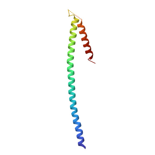
    4E61

  • PubMed Abstract: 

    Mitosis begins with the tethering of chromosomes to the mitotic spindle and their orientation perpendicular to the axis of cell division. In budding yeast, mitotic spindle orientation and the subsequent chromosome segregation are two independent processes. Early spindle orientation is driven by the actin-bound myosin Myo2p, which interacts with the adapter Kar9p. The latter also binds to microtubule-associated Bim1p, thereby connecting both types of cytoskeleton. This study focuses on the interaction between Kar9p and Bim1p and its regulation. We solved the crystal structure of the previously reported Kar9p-binding motif of Bim1p and identified a second, novel Kar9p interaction domain. We further show that two independent post-translational modification events regulate their interaction. Whereas Kar9p sumoylation is required for efficient complex formation with Bim1p, Aurora B/Ipl1p-dependent phosphorylation of Bim1p down-regulates their interaction. The observed effects of these modifications allow us to propose a novel regulatory framework for the assembly and disassembly of the early spindle orientation complex.


  • Organizational Affiliation
    • Institute of Structural Biology, Helmholtz Zentrum München-German Research Center for Environmental Health, 85764 Neuherberg, Germany.

Macromolecule Content 

  • Total Structure Weight: 23.62 kDa 
  • Atom Count: 1,363 
  • Modeled Residue Count: 175 
  • Deposited Residue Count: 212 
  • Unique protein chains: 1

Macromolecules

Find similar proteins by:|  3D Structure
Entity ID: 1
MoleculeChains  Sequence LengthOrganismDetailsImage
Protein BIM1
A, B
106Saccharomyces cerevisiaeMutation(s): 0 
Gene Names: BIM1YER016W
UniProt
Find proteins for P40013 (Saccharomyces cerevisiae (strain ATCC 204508 / S288c))
Explore P40013 
Go to UniProtKB:  P40013
Entity Groups
Sequence Clusters30% Identity50% Identity70% Identity90% Identity95% Identity100% Identity
UniProt GroupP40013
Sequence Annotations
Expand
Reference Sequence

Experimental Data & Validation

Experimental Data

  • Method: X-RAY DIFFRACTION
  • Resolution: 2.45 Å
  • R-Value Free:  0.241 (Depositor), 0.271 (DCC) 
  • R-Value Work:  0.211 (Depositor), 0.226 (DCC) 
Space Group: P 1 21 1
Unit Cell:
Length ( Å )Angle ( ˚ )
a = 28α = 90
b = 42.5β = 90.3
c = 100.5γ = 90
Software Package:
Software NamePurpose
XCUBEdata collection
SHELXmodel building
SHELXDphasing
SHELXEmodel building
BUSTER-TNTrefinement
XDSdata reduction
XSCALEdata scaling
SHELXphasing

Structure Validation

View Full Validation Report


Entry History 

Deposition Data

Revision History  (Full details and data files)

  • Version 1.0: 2012-04-11
    Type: Initial release
  • Version 1.1: 2012-06-13
    Changes: Database references
  • Version 1.2: 2017-11-15
    Changes: Refinement description
  • Version 1.3: 2024-02-28
    Changes: Data collection, Database references
  • Version 1.4: 2024-04-03
    Changes: Refinement description