4E2F | pdb_00004e2f

Crystal Structure of E. coli Aspartate Transcarbamoylase K164E/E239K Mutant in an intermediate state


Experimental Data Snapshot

  • Method: X-RAY DIFFRACTION
  • Resolution: 2.80 Å
  • R-Value Free: 
    0.274 (Depositor), 0.268 (DCC) 
  • R-Value Work: 
    0.214 (Depositor), 0.207 (DCC) 
  • R-Value Observed: 
    0.217 (Depositor) 

wwPDB Validation 3D Report Full Report

Validation slider image for 4E2F

This is version 1.3 of the entry. See complete history

Literature

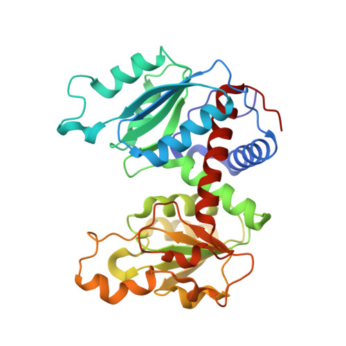
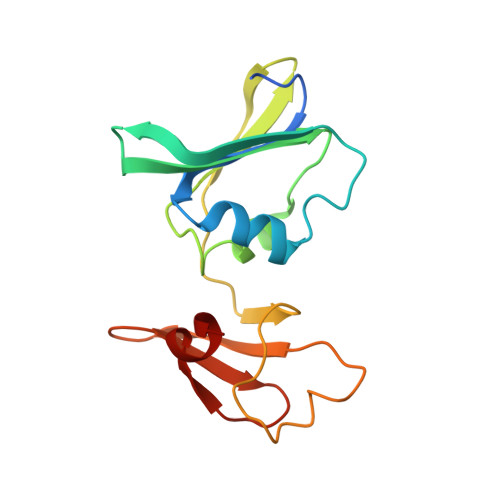
Trapping and structure determination of an intermediate in the allosteric transition of aspartate transcarbamoylase.

Guo, W.West, J.M.Dutton, A.S.Tsuruta, H.Kantrowitz, E.R.

(2012) Proc Natl Acad Sci U S A 109: 7741-7746

  • DOI: https://doi.org/10.1073/pnas.1119683109
  • Primary Citation Related Structures: 
    4E2F

  • PubMed Abstract: 

    X-ray crystallography and small-angle X-ray scattering (SAXS) in solution have been used to show that a mutant aspartate transcarbamoylase exists in an intermediate quaternary structure between the canonical T and R structures. Additionally, the SAXS data indicate a pH-dependent structural alteration consistent with either a pH-induced conformational change or a pH-induced alteration in the T to R equilibrium. These data indicate that this mutant is not a model for the R state, as has been proposed, but rather represents the enzyme trapped along the path of the allosteric transition between the T and R states.


  • Organizational Affiliation
    • Department of Chemistry, Merkert Chemistry Center, Boston College, Chestnut Hill, MA 02467, USA.

Macromolecule Content 

  • Total Structure Weight: 309.28 kDa 
  • Atom Count: 21,764 
  • Modeled Residue Count: 2,724 
  • Deposited Residue Count: 2,778 
  • Unique protein chains: 2

Macromolecules

Find similar proteins by:|  3D Structure
Entity ID: 1
MoleculeChains  Sequence LengthOrganismDetailsImage
Aspartate carbamoyltransferase catalytic chain310Escherichia coli K-12Mutation(s): 2 
Gene Names: b4245JW4204pyrB
EC: 2.1.3.2
UniProt
Find proteins for P0A786 (Escherichia coli (strain K12))
Explore P0A786 
Go to UniProtKB:  P0A786
Entity Groups
Sequence Clusters30% Identity50% Identity70% Identity90% Identity95% Identity100% Identity
UniProt GroupP0A786
Sequence Annotations
Expand
Reference Sequence
Find similar proteins by:|  3D Structure
Entity ID: 2
MoleculeChains  Sequence LengthOrganismDetailsImage
Aspartate carbamoyltransferase regulatory chain153Escherichia coli K-12Mutation(s): 0 
Gene Names: b4244JW4203pyrI
UniProt
Find proteins for P0A7F3 (Escherichia coli (strain K12))
Explore P0A7F3 
Go to UniProtKB:  P0A7F3
Entity Groups
Sequence Clusters30% Identity50% Identity70% Identity90% Identity95% Identity100% Identity
UniProt GroupP0A7F3
Sequence Annotations
Expand
Reference Sequence

Experimental Data & Validation

Experimental Data

  • Method: X-RAY DIFFRACTION
  • Resolution: 2.80 Å
  • R-Value Free:  0.274 (Depositor), 0.268 (DCC) 
  • R-Value Work:  0.214 (Depositor), 0.207 (DCC) 
  • R-Value Observed: 0.217 (Depositor) 
Space Group: P 21 21 21
Unit Cell:
Length ( Å )Angle ( ˚ )
a = 124.086α = 90
b = 144.814β = 90
c = 203.359γ = 90
Software Package:
Software NamePurpose
CrystalCleardata collection
MOLREPphasing
PHENIXrefinement
d*TREKdata reduction
d*TREKdata scaling

Structure Validation

View Full Validation Report


Entry History 

Deposition Data

Revision History  (Full details and data files)

  • Version 1.0: 2012-05-02
    Type: Initial release
  • Version 1.1: 2012-05-16
    Changes: Database references
  • Version 1.2: 2012-05-30
    Changes: Database references
  • Version 1.3: 2024-02-28
    Changes: Data collection, Database references, Derived calculations, Refinement description