4DWX | pdb_00004dwx

Crystal Structure of a Family GH-19 Chitinase from rye seeds


Experimental Data Snapshot

  • Method: X-RAY DIFFRACTION
  • Resolution: 1.80 Å
  • R-Value Free: 
    0.204 (Depositor), 0.203 (DCC) 
  • R-Value Work: 
    0.179 (Depositor), 0.179 (DCC) 
  • R-Value Observed: 
    0.179 (Depositor) 

Starting Model: experimental
View more details

wwPDB Validation 3D Report Full Report

Validation slider image for 4DWX

This is version 1.3 of the entry. See complete history

Literature

Crystal structure and chitin oligosaccharide-binding mode of a 'loopful' family GH19 chitinase from rye, Secale cereale, seeds

Ohnuma, T.Numata, T.Osawa, T.Inanaga, H.Okazaki, Y.Shinya, S.Kondo, K.Fukuda, T.Fukamizo, T.

(2012) FEBS J 279: 3639-3651

  • DOI: https://doi.org/10.1111/j.1742-4658.2012.08723.x
  • Primary Citation Related Structures: 
    4DWX, 4DYG

  • PubMed Abstract: 

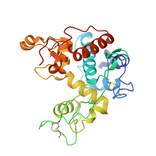
    The substrate-binding mode of a 26-kDa GH19 chitinase from rye, Secale cereale, seeds (RSC-c) was investigated by crystallography, site-directed mutagenesis and NMR spectroscopy. The crystal structure of RSC-c in a complex with an N-acetylglucosamine tetramer, (GlcNAc)(4) , was successfully solved, and revealed the binding mode of the tetramer to be an aglycon-binding site, subsites +1, +2, +3, and +4. These are the first crystallographic data showing the oligosaccharide-binding mode of a family GH19 chitinase. From HPLC analysis of the enzymatic reaction products, mutation of Trp72 to alanine was found to affect the product distribution obtained from the substrate, p-nitrophenyl penta-N-acetyl-β-chitopentaoside. Mutational experiments confirmed the crystallographic finding that the Trp72 side chain interacts with the +4 moiety of the bound substrate. To further confirm the crystallographic data, binding experiments were also conducted in solution using NMR spectroscopy. Several signals in the (1) H-(15) N HSQC spectrum of the stable isotope-labeled RSC-c were affected upon addition of (GlcNAc)(4) . Signal assignments revealed that most signals responsive to the addition of (GlcNAc)(4) are derived from amino acids located at the surface of the aglycon-binding site. The binding mode deduced from NMR binding experiments in solution was consistent with that from the crystal structure.


  • Organizational Affiliation
    •  Department of Advanced Bioscience, Kinki University, Nara, Japan Biomedical Research Institute, National Institute of Advanced Industrial Science and Technology (AIST), Tsukuba, Japan.

Macromolecule Content 

  • Total Structure Weight: 53.52 kDa 
  • Atom Count: 4,281 
  • Modeled Residue Count: 484 
  • Deposited Residue Count: 488 
  • Unique protein chains: 1

Macromolecules

Find similar proteins by:|  3D Structure
Entity ID: 1
MoleculeChains  Sequence LengthOrganismDetailsImage
Basic endochitinase C
A, B
244Secale cerealeMutation(s): 0 
Gene Names: rscc
EC: 3.2.1.14
UniProt
Find proteins for Q9FRV0 (Secale cereale)
Explore Q9FRV0 
Go to UniProtKB:  Q9FRV0
Entity Groups
Sequence Clusters30% Identity50% Identity70% Identity90% Identity95% Identity100% Identity
UniProt GroupQ9FRV0
Sequence Annotations
Expand
Reference Sequence

Small Molecules

Ligands 3 Unique
IDChains Name / Formula / InChI Key2D Diagram3D Interactions
MES

Query on MES


Download:Ideal Coordinates CCD File
C [auth A],
J [auth B]
2-(N-MORPHOLINO)-ETHANESULFONIC ACID
C6 H13 N O4 S
SXGZJKUKBWWHRA-UHFFFAOYSA-N
SO4

Query on SO4


Download:Ideal Coordinates CCD File
D [auth A],
E [auth A],
K [auth B]
SULFATE ION
O4 S
QAOWNCQODCNURD-UHFFFAOYSA-L
ZN

Query on ZN


Download:Ideal Coordinates CCD File
F [auth A],
G [auth A],
H [auth A],
I [auth A],
L [auth B]
ZINC ION
Zn
PTFCDOFLOPIGGS-UHFFFAOYSA-N

Experimental Data & Validation

Experimental Data

  • Method: X-RAY DIFFRACTION
  • Resolution: 1.80 Å
  • R-Value Free:  0.204 (Depositor), 0.203 (DCC) 
  • R-Value Work:  0.179 (Depositor), 0.179 (DCC) 
  • R-Value Observed: 0.179 (Depositor) 
Space Group: P 21 21 21
Unit Cell:
Length ( Å )Angle ( ˚ )
a = 64.914α = 90
b = 84.684β = 90
c = 108.815γ = 90
Software Package:
Software NamePurpose
HKL-2000data collection
PHASERphasing
CNSrefinement
HKL-2000data reduction
HKL-2000data scaling

Structure Validation

View Full Validation Report


Entry History 

Deposition Data

Revision History  (Full details and data files)

  • Version 1.0: 2012-08-15
    Type: Initial release
  • Version 1.1: 2013-06-26
    Changes: Database references
  • Version 1.2: 2023-11-08
    Changes: Data collection, Database references, Derived calculations, Refinement description
  • Version 1.3: 2024-10-30
    Changes: Structure summary