4DVI

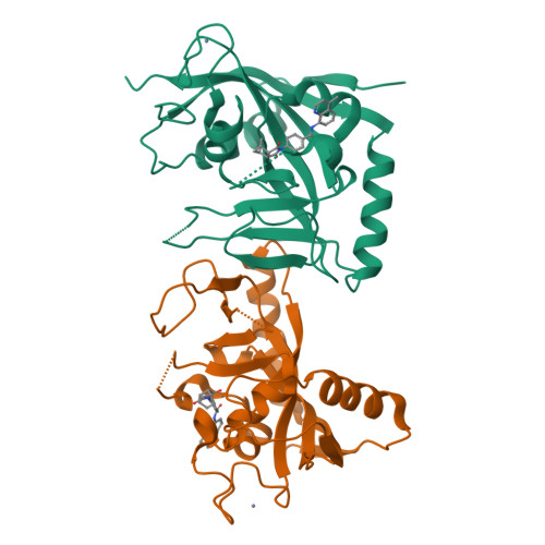
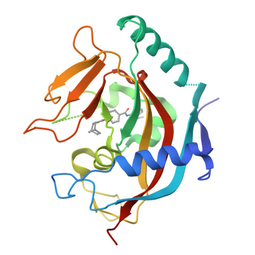
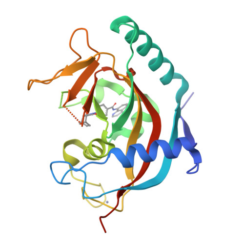
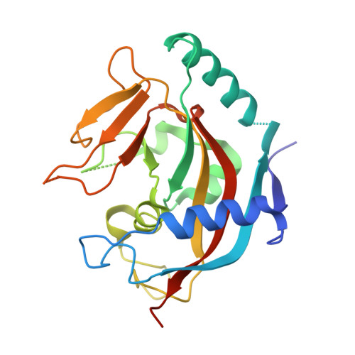
Crystal structure of Tankyrase 1 with IWR2


Experimental Data Snapshot

  • Method: X-RAY DIFFRACTION
  • Resolution: 1.90 Å
  • R-Value Free: 0.258 
  • R-Value Work: 0.248 

wwPDB Validation   3D Report Full Report


Ligand Structure Quality Assessment 


This is version 1.1 of the entry. See complete history


Literature

Novel binding mode of a potent and selective tankyrase inhibitor.

Gunaydin, H.Gu, Y.Huang, X.

(2012) PLoS One 7: e33740-e33740

  • DOI: https://doi.org/10.1371/journal.pone.0033740
  • Primary Citation of Related Structures:  
    4DVI

  • PubMed Abstract: 

    Tankyrases (TNKS1 and TNKS2) are key regulators of cellular processes such as telomere pathway and Wnt signaling. IWRs (inhibitors of Wnt response) have recently been identified as potent and selective inhibitors of tankyrases. However, it is not clear how these IWRs interact with tankyrases. Here we report the crystal structure of the catalytic domain of human TNKS1 in complex with IWR2, which reveals a novel binding site for tankyrase inhibitors. The TNKS1/IWR2 complex provides a molecular basis for their strong and specific interactions and suggests clues for further development of tankyrase inhibitors.


  • Organizational Affiliation

    Department of Molecular Structure, Amgen, Cambridge, Massachusetts, United States of America.


Macromolecules
Find similar proteins by:  (by identity cutoff)  |  3D Structure
Entity ID: 1
MoleculeChains Sequence LengthOrganismDetailsImage
Tankyrase-1
A, B
217Homo sapiensMutation(s): 0 
Gene Names: TNKSPARP5APARPLTIN1TINF1TNKS1
EC: 2.4.2.30 (PDB Primary Data), 2.4.2 (UniProt)
UniProt & NIH Common Fund Data Resources
Find proteins for O95271 (Homo sapiens)
Explore O95271 
Go to UniProtKB:  O95271
PHAROS:  O95271
GTEx:  ENSG00000173273 
Entity Groups  
Sequence Clusters30% Identity50% Identity70% Identity90% Identity95% Identity100% Identity
UniProt GroupO95271
Sequence Annotations
Expand
  • Reference Sequence
Small Molecules
Experimental Data & Validation

Experimental Data

  • Method: X-RAY DIFFRACTION
  • Resolution: 1.90 Å
  • R-Value Free: 0.258 
  • R-Value Work: 0.248 
  • Space Group: P 21 21 21
Unit Cell:
Length ( Å )Angle ( ˚ )
a = 41.47α = 90
b = 77.94β = 90
c = 146.54γ = 90
Software Package:
Software NamePurpose
HKL-2000data collection
AMoREphasing
CNSrefinement
HKL-2000data reduction
HKL-2000data scaling

Structure Validation

View Full Validation Report


Ligand Structure Quality Assessment 


Entry History 

Deposition Data

  • Released Date: 2012-04-04 
  • Deposition Author(s): Huang, X.

Revision History  (Full details and data files)

  • Version 1.0: 2012-04-04
    Type: Initial release
  • Version 1.1: 2024-02-28
    Changes: Data collection, Database references, Derived calculations