4DUX | pdb_00004dux

E. coli (lacZ) beta-galactosidase (N460S) in complex with L-ribose


Experimental Data Snapshot

  • Method: X-RAY DIFFRACTION
  • Resolution: 2.30 Å
  • R-Value Free: 
    0.221 (Depositor), 0.223 (DCC) 
  • R-Value Work: 
    0.170 (Depositor), 0.173 (DCC) 
  • R-Value Observed: 
    0.170 (Depositor) 

Starting Model: experimental
View more details

wwPDB Validation 3D Report Full Report

Validation slider image for 4DUX

Ligand Structure Quality Assessment 


This is version 1.4 of the entry. See complete history

Literature

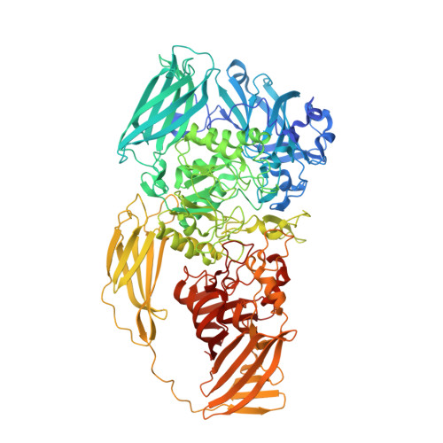
Structural explanation for allolactose (lac operon inducer) synthesis by lacZ beta-galactosidase and the evolutionary relationship between allolactose synthesis and the lac repressor.

Wheatley, R.W.Lo, S.Jancewicz, L.J.Dugdale, M.L.Huber, R.E.

(2013) J Biological Chem 288: 12993-13005

  • DOI: https://doi.org/10.1074/jbc.M113.455436
  • Primary Citation Related Structures: 
    4DUW, 4DUX

  • PubMed Abstract: 

    β-Galactosidase (lacZ) has bifunctional activity. It hydrolyzes lactose to galactose and glucose and catalyzes the intramolecular isomerization of lactose to allolactose, the lac operon inducer. β-Galactosidase promotes the isomerization by means of an acceptor site that binds glucose after its cleavage from lactose and thus delays its exit from the site. However, because of its relatively low affinity for glucose, details of this site have remained elusive. We present structural data mapping the glucose site based on a substituted enzyme (G794A-β-galactosidase) that traps allolactose. Various lines of evidence indicate that the glucose of the trapped allolactose is in the acceptor position. The evidence includes structures with Bis-Tris (2,2-bis(hydroxymethyl)-2,2',2″-nitrilotriethanol) and L-ribose in the site and kinetic binding studies with substituted β-galactosidases. The site is composed of Asn-102, His-418, Lys-517, Ser-796, Glu-797, and Trp-999. Ser-796 and Glu-797 are part of a loop (residues 795-803) that closes over the active site. This loop appears essential for the bifunctional nature of the enzyme because it helps form the glucose binding site. In addition, because the loop is mobile, glucose binding is transient, allowing the release of some glucose. Bioinformatics studies showed that the residues important for interacting with glucose are only conserved in a subset of related enzymes. Thus, intramolecular isomerization is not a universal feature of β-galactosidases. Genomic analyses indicated that lac repressors were co-selected only within the conserved subset. This shows that the glucose binding site of β-galactosidase played an important role in lac operon evolution.


  • Organizational Affiliation
    • Division of Biochemistry, Faculty of Science, University of Calgary, Calgary, Alberta T2N 1N4, Canada.

Macromolecule Content 

  • Total Structure Weight: 485.81 kDa 
  • Atom Count: 35,621 
  • Modeled Residue Count: 4,060 
  • Deposited Residue Count: 4,208 
  • Unique protein chains: 1

Macromolecules

Find similar proteins by:|  3D Structure
Entity ID: 1
MoleculeChains  Sequence LengthOrganismDetailsImage
Beta-galactosidase
A, B, C, D
1,052Escherichia coli K-12Mutation(s): 1 
Gene Names: b0344JW0335lacZ
EC: 3.2.1.23
UniProt
Find proteins for P00722 (Escherichia coli (strain K12))
Explore P00722 
Go to UniProtKB:  P00722
Entity Groups
Sequence Clusters30% Identity50% Identity70% Identity90% Identity95% Identity100% Identity
UniProt GroupP00722
Sequence Annotations
Expand
Reference Sequence

Small Molecules

Ligands 4 Unique
IDChains Name / Formula / InChI Key2D Diagram3D Interactions
0MK

Query on 0MK


Download:Ideal Coordinates CCD File
AB [auth C]
BB [auth C]
DA [auth B]
E [auth A]
EA [auth B]
AB [auth C],
BB [auth C],
DA [auth B],
E [auth A],
EA [auth B],
F [auth A],
FA [auth B],
XB [auth D],
YB [auth D]
beta-L-ribopyranose
C5 H10 O5
SRBFZHDQGSBBOR-FCAWWPLPSA-N
DMS

Query on DMS


Download:Ideal Coordinates CCD File
AA [auth A]
BA [auth A]
CA [auth A]
EC [auth D]
FC [auth D]
AA [auth A],
BA [auth A],
CA [auth A],
EC [auth D],
FC [auth D],
GC [auth D],
HC [auth D],
IB [auth C],
IC [auth D],
JB [auth C],
JC [auth D],
KB [auth C],
KC [auth D],
L [auth A],
LB [auth C],
LC [auth D],
M [auth A],
MA [auth B],
MB [auth C],
MC [auth D],
N [auth A],
NA [auth B],
NB [auth C],
NC [auth D],
O [auth A],
OA [auth B],
OB [auth C],
OC [auth D],
P [auth A],
PA [auth B],
PB [auth C],
PC [auth D],
Q [auth A],
QA [auth B],
QB [auth C],
QC [auth D],
R [auth A],
RA [auth B],
RB [auth C],
RC [auth D],
S [auth A],
SA [auth B],
SB [auth C],
SC [auth D],
T [auth A],
TA [auth B],
TB [auth C],
TC [auth D],
U [auth A],
UA [auth B],
UB [auth C],
UC [auth D],
V [auth A],
VA [auth B],
VB [auth C],
VC [auth D],
W [auth A],
WA [auth B],
WB [auth C],
X [auth A],
XA [auth B],
Y [auth A],
YA [auth B],
Z [auth A],
ZA [auth B]
DIMETHYL SULFOXIDE
C2 H6 O S
IAZDPXIOMUYVGZ-UHFFFAOYSA-N
MG

Query on MG


Download:Ideal Coordinates CCD File
AC [auth D]
CB [auth C]
DB [auth C]
G [auth A]
GA [auth B]
AC [auth D],
CB [auth C],
DB [auth C],
G [auth A],
GA [auth B],
H [auth A],
HA [auth B],
ZB [auth D]
MAGNESIUM ION
Mg
JLVVSXFLKOJNIY-UHFFFAOYSA-N
NA

Query on NA


Download:Ideal Coordinates CCD File
BC [auth D]
CC [auth D]
DC [auth D]
EB [auth C]
FB [auth C]
BC [auth D],
CC [auth D],
DC [auth D],
EB [auth C],
FB [auth C],
GB [auth C],
HB [auth C],
I [auth A],
IA [auth B],
J [auth A],
JA [auth B],
K [auth A],
KA [auth B],
LA [auth B]
SODIUM ION
Na
FKNQFGJONOIPTF-UHFFFAOYSA-N

Experimental Data & Validation

Experimental Data

  • Method: X-RAY DIFFRACTION
  • Resolution: 2.30 Å
  • R-Value Free:  0.221 (Depositor), 0.223 (DCC) 
  • R-Value Work:  0.170 (Depositor), 0.173 (DCC) 
  • R-Value Observed: 0.170 (Depositor) 
Space Group: P 21 21 21
Unit Cell:
Length ( Å )Angle ( ˚ )
a = 150.648α = 90
b = 168.103β = 90
c = 201.881γ = 90
Software Package:
Software NamePurpose
REFMACrefinement
CNSrefinement
ADSCdata collection
MOSFLMdata reduction
SCALEPACKdata scaling
CNSphasing

Structure Validation

View Full Validation Report


Ligand Structure Quality Assessment 


Entry History 

Deposition Data

Revision History  (Full details and data files)

  • Version 1.0: 2013-03-20
    Type: Initial release
  • Version 1.1: 2013-09-04
    Changes: Database references
  • Version 1.2: 2019-07-17
    Changes: Data collection, Refinement description
  • Version 1.3: 2020-07-29
    Type: Remediation
    Reason: Carbohydrate remediation
    Changes: Data collection, Database references, Derived calculations, Structure summary
  • Version 1.4: 2023-09-13
    Changes: Data collection, Database references, Refinement description, Structure summary