4DUK | pdb_00004duk

Carboxypeptidase T with L-BENZYLSUCCINIC ACID


Experimental Data Snapshot

  • Method: X-RAY DIFFRACTION
  • Resolution: 1.57 Å
  • R-Value Free: 
    0.167 (Depositor), 0.167 (DCC) 
  • R-Value Work: 
    0.151 (Depositor), 0.150 (DCC) 
  • R-Value Observed: 
    0.152 (Depositor) 

wwPDB Validation 3D Report Full Report

Validation slider image for 4DUK

Ligand Structure Quality Assessment 


This is version 1.2 of the entry. See complete history

Literature

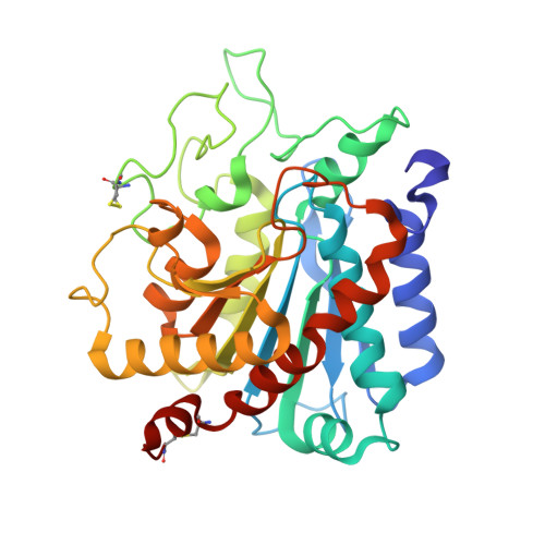
Structural insights into the broad substrate specificity of carboxypeptidase T from Thermoactinomyces vulgaris.

Akparov, V.K.h.Timofeev, V.I.Khaliullin, I.G.Svedas, V.Chestukhina, G.G.Kuranova, I.P.

(2015) FEBS J 282: 1214-1224

  • DOI: https://doi.org/10.1111/febs.13210
  • Primary Citation Related Structures: 
    3V7Z, 4DUK

  • PubMed Abstract: 

    The crystal structures of carboxypeptidase T (CpT) complexes with phenylalanine and arginine substrate analogs - benzylsuccinic acid and (2-guanidinoethylmercapto)succinic acid - were determined by the molecular replacement method at resolutions of 1.57 Å and 1.62 Å to clarify the broad substrate specificity profile of the enzyme. The conservative Leu211 and Leu254 residues (also present in both carboxypeptidase A and carboxypeptidase B) were shown to be structural determinants for recognition of hydrophobic substrates, whereas Asp263 was for recognition of positively charged substrates. Mutations of these determinants modify the substrate profile: the CpT variant Leu211Gln acquires carboxypeptidase B-like properties, and the CpT variant Asp263Asn the carboxypeptidase A-like selectivity. The Pro248-Asp258 loop interacting with Leu254 and Tyr255 was shown to be responsible for recognition of the substrate's C-terminal residue. Substrate binding at the S1' subsite leads to the ligand-dependent shift of this loop, and Leu254 side chain movement induces the conformation rearrangement of the Glu277 residue crucial for catalysis. This is a novel insight into the substrate selectivity of metallocarboxypeptidases that demonstrates the importance of interactions between the S1' subsite and the catalytic center.


  • Organizational Affiliation
    • State Research Institute for Genetics and Selection of Industrial Microorganisms, Moscow, Russia.

Macromolecule Content 

  • Total Structure Weight: 39.79 kDa 
  • Atom Count: 3,298 
  • Modeled Residue Count: 323 
  • Deposited Residue Count: 326 
  • Unique protein chains: 1

Macromolecules

Find similar proteins by:|  3D Structure
Entity ID: 1
MoleculeChains  Sequence LengthOrganismDetailsImage
Carboxypeptidase T326Thermoactinomyces vulgarisMutation(s): 0 
Gene Names: cpt
EC: 3.4.17.18
UniProt
Find proteins for P29068 (Thermoactinomyces vulgaris)
Explore P29068 
Go to UniProtKB:  P29068
Entity Groups
Sequence Clusters30% Identity50% Identity70% Identity90% Identity95% Identity100% Identity
UniProt GroupP29068
Sequence Annotations
Expand
Reference Sequence

Small Molecules

Ligands 5 Unique
IDChains Name / Formula / InChI Key2D Diagram3D Interactions
BZS

Query on BZS


Download:Ideal Coordinates CCD File
F [auth A]L-BENZYLSUCCINIC ACID
C11 H12 O4
GTOFKXZQQDSVFH-SECBINFHSA-N
SO4

Query on SO4


Download:Ideal Coordinates CCD File
CA [auth A],
G [auth A]
SULFATE ION
O4 S
QAOWNCQODCNURD-UHFFFAOYSA-L
GOL

Query on GOL


Download:Ideal Coordinates CCD File
AA [auth A]
DA [auth A]
EA [auth A]
FA [auth A]
GA [auth A]
AA [auth A],
DA [auth A],
EA [auth A],
FA [auth A],
GA [auth A],
H [auth A],
I [auth A],
J [auth A],
K [auth A],
L [auth A],
M [auth A],
N [auth A],
O [auth A],
P [auth A],
Q [auth A],
R [auth A],
S [auth A],
T [auth A],
U [auth A],
V [auth A],
W [auth A],
X [auth A],
Y [auth A],
Z [auth A]
GLYCEROL
C3 H8 O3
PEDCQBHIVMGVHV-UHFFFAOYSA-N
ZN

Query on ZN


Download:Ideal Coordinates CCD File
B [auth A]ZINC ION
Zn
PTFCDOFLOPIGGS-UHFFFAOYSA-N
CA

Query on CA


Download:Ideal Coordinates CCD File
BA [auth A],
C [auth A],
D [auth A],
E [auth A]
CALCIUM ION
Ca
BHPQYMZQTOCNFJ-UHFFFAOYSA-N

Experimental Data & Validation

Experimental Data

  • Method: X-RAY DIFFRACTION
  • Resolution: 1.57 Å
  • R-Value Free:  0.167 (Depositor), 0.167 (DCC) 
  • R-Value Work:  0.151 (Depositor), 0.150 (DCC) 
  • R-Value Observed: 0.152 (Depositor) 
Space Group: P 63 2 2
Unit Cell:
Length ( Å )Angle ( ˚ )
a = 157.939α = 90
b = 157.939β = 90
c = 104.437γ = 120
Software Package:
Software NamePurpose
HKL-2000data collection
PHASERphasing
REFMACrefinement
HKL-2000data reduction
HKL-2000data scaling

Structure Validation

View Full Validation Report


Ligand Structure Quality Assessment 


Entry History 

Deposition Data

Revision History  (Full details and data files)

  • Version 1.0: 2013-02-27
    Type: Initial release
  • Version 1.1: 2015-09-30
    Changes: Database references
  • Version 1.2: 2024-11-20
    Changes: Data collection, Database references, Derived calculations, Experimental preparation, Structure summary