


4DO0 | pdb_00004do0
4DO0 | pdb_00004do0
Crystal Structure of human PHF8 in complex with Daminozide
- PDB DOI: https://doi.org/10.2210/pdb4DO0/pdb
- Classification: METAL BINDING PROTEIN
- Organism(s): Homo sapiens
- Expression System: Escherichia coli
- Mutation(s): No
- Deposited: 2012-02-09 Released: 2012-03-14
Experimental Data Snapshot
- Method: X-RAY DIFFRACTION
- Resolution: 2.55 Å
- R-Value Free: 0.265 (Depositor), 0.253 (DCC)
- R-Value Work: 0.208 (Depositor)
- R-Value Observed: 0.211 (Depositor)
Starting Model: experimental
View more details

This is version 1.2 of the entry. See complete history.

Macromolecules
Find similar proteins by:
| 3D Structure
Entity ID: 1 | |||||
|---|---|---|---|---|---|
| Molecule | Chains | Sequence Length | Organism | Details | Image |
| Histone lysine demethylase PHF8 | 374 | Homo sapiens | Mutation(s): 0 Gene Names: PHF8, KIAA1111, ZNF422 EC: 1.14.11 (UniProt), 1.14.11.65 (UniProt) | 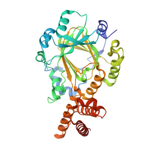 | |
UniProt & NIH Common Fund Data Resources | |||||
PHAROS: Q9UPP1 GTEx: ENSG00000172943 | |||||
Entity Groups | |||||
| Sequence Clusters | 30% Identity50% Identity70% Identity90% Identity95% Identity100% Identity | ||||
| UniProt Group | Q9UPP1 | ||||
Sequence AnnotationsExpand | |||||
Reference Sequence | |||||
Small Molecules
| Ligands 5 Unique | |||||
|---|---|---|---|---|---|
| ID | Chains | Name / Formula / InChI Key | 2D Diagram | 3D Interactions | |
| DZA Download:Ideal Coordinates CCD File | B [auth A] | DAMINOZIDE C6 H12 N2 O3 NOQGZXFMHARMLW-UHFFFAOYSA-N |  | ||
| SO4 Download:Ideal Coordinates CCD File | D [auth A] E [auth A] F [auth A] G [auth A] H [auth A] | SULFATE ION O4 S QAOWNCQODCNURD-UHFFFAOYSA-L |  | ||
| ZN Download:Ideal Coordinates CCD File | C [auth A] | ZINC ION Zn PTFCDOFLOPIGGS-UHFFFAOYSA-N |  | ||
| EDO Download:Ideal Coordinates CCD File | J [auth A], L [auth A], M [auth A] | 1,2-ETHANEDIOL C2 H6 O2 LYCAIKOWRPUZTN-UHFFFAOYSA-N |  | ||
| ACT Download:Ideal Coordinates CCD File | K [auth A], N [auth A] | ACETATE ION C2 H3 O2 QTBSBXVTEAMEQO-UHFFFAOYSA-M |  | ||
Experimental Data & Validation
Experimental Data
- Method: X-RAY DIFFRACTION
- Resolution: 2.55 Å
- R-Value Free: 0.265 (Depositor), 0.253 (DCC)
- R-Value Work: 0.208 (Depositor)
- R-Value Observed: 0.211 (Depositor)
Space Group: I 21 3
Unit Cell:
| Length ( Å ) | Angle ( ˚ ) |
|---|---|
| a = 151.734 | α = 90 |
| b = 151.734 | β = 90 |
| c = 151.734 | γ = 90 |
| Software Name | Purpose |
|---|---|
| GDA | data collection |
| PHASER | phasing |
| REFMAC | refinement |
| XDS | data reduction |
| SCALA | data scaling |
Entry History
Deposition Data
- Released Date: 2012-03-14 Deposition Author(s): Krojer, T., Daniel, M., Ng, S.S., Walport, L.J., Chowdhury, R., Arrowsmith, C.H., Edwards, A., Bountra, C., Kawamura, A., Muller-Knapp, S., McDonough, M.A., von Delft, F., Schofield, C.J., Oppermann, U., Structural Genomics Consortium (SGC)
Revision History (Full details and data files)
- Version 1.0: 2012-03-14
Type: Initial release - Version 1.1: 2020-02-26
Changes: Data collection, Database references - Version 1.2: 2023-09-13
Changes: Data collection, Database references, Derived calculations, Refinement description


