4DK4 | pdb_00004dk4

Crystal Structure of Trypanosoma brucei dUTPase with dUpNp, Ca2+ and Na+


Experimental Data Snapshot

  • Method: X-RAY DIFFRACTION
  • Resolution: 1.90 Å
  • R-Value Free: 
    0.232 (Depositor), 0.235 (DCC) 
  • R-Value Work: 
    0.177 (Depositor), 0.182 (DCC) 
  • R-Value Observed: 
    0.180 (Depositor) 

Starting Model: experimental
View more details

wwPDB Validation 3D Report Full Report

Validation slider image for 4DK4

Ligand Structure Quality Assessment 


This is version 1.3 of the entry. See complete history

Literature

On the catalytic mechanism of dimeric dUTPases.

Hemsworth, G.R.Gonzalez-Pacanowska, D.Wilson, K.S.

(2013) Biochem J 456: 81-88

  • DOI: https://doi.org/10.1042/BJ20130796
  • Primary Citation Related Structures: 
    4DK2, 4DK4, 4DKB, 4DL8, 4DLC

  • PubMed Abstract: 

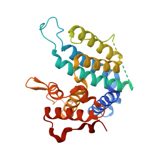
    The Tritryps Trypanosoma brucei, Trypanosoma cruzi and Leishmania donovani are responsible for great morbidity and mortality in developing countries. Their dimeric dUTPases are members of the all-α NTP pyrophosphohydrolase family and represent promising drug targets due to their essential nature and markedly different structural and biochemical properties compared with the trimeric human enzyme. In the present paper we describe the structure of the T. brucei enzyme in open and closed conformations. Furthermore, we probe the reaction mechanism through the binding of transition state mimics both in solution and in the crystal. 31P-NMR and tryptophan fluorescence quenching in the presence of AlF3 and MgF3- identified which phosphate is subject to nucleophilic attack by a water molecule. The structures in complex with two transition state analogues confirm that the nucleophilic attack occurs on the β-phosphate in contrast with the α-phosphate in the trimeric enzymes. These results establish the structural basis of catalysis of these important housekeeping enzymes and has ramifications for the wider all-α NTP pyrophosphohydrolase family.


  • Organizational Affiliation
    • *Structural Biology Laboratory, Department of Chemistry, University of York, Heslington, York YO10 5DD, U.K.

Macromolecule Content 

  • Total Structure Weight: 65.39 kDa 
  • Atom Count: 3,562 
  • Modeled Residue Count: 435 
  • Deposited Residue Count: 580 
  • Unique protein chains: 1

Macromolecules

Find similar proteins by:|  3D Structure
Entity ID: 1
MoleculeChains  Sequence LengthOrganismDetailsImage
Deoxyuridine triphosphatase
A, B
290Trypanosoma bruceiMutation(s): 0 
Gene Names: Tb927.7.5160
EC: 3.6.1.23
UniProt
Find proteins for Q57ZH3 (Trypanosoma brucei brucei (strain 927/4 GUTat10.1))
Explore Q57ZH3 
Go to UniProtKB:  Q57ZH3
Entity Groups
Sequence Clusters30% Identity50% Identity70% Identity90% Identity95% Identity100% Identity
UniProt GroupQ57ZH3
Sequence Annotations
Expand
Reference Sequence

Small Molecules

Ligands 3 Unique
IDChains Name / Formula / InChI Key2D Diagram3D Interactions
DUN

Query on DUN


Download:Ideal Coordinates CCD File
C [auth A],
G [auth B]
2'-DEOXYURIDINE 5'-ALPHA,BETA-IMIDO-DIPHOSPHATE
C9 H15 N3 O10 P2
COFNIXBQVWFHTR-SHYZEUOFSA-N
CA

Query on CA


Download:Ideal Coordinates CCD File
D [auth A],
E [auth A],
H [auth B],
I [auth B]
CALCIUM ION
Ca
BHPQYMZQTOCNFJ-UHFFFAOYSA-N
NA

Query on NA


Download:Ideal Coordinates CCD File
F [auth A],
J [auth B]
SODIUM ION
Na
FKNQFGJONOIPTF-UHFFFAOYSA-N

Experimental Data & Validation

Experimental Data

  • Method: X-RAY DIFFRACTION
  • Resolution: 1.90 Å
  • R-Value Free:  0.232 (Depositor), 0.235 (DCC) 
  • R-Value Work:  0.177 (Depositor), 0.182 (DCC) 
  • R-Value Observed: 0.180 (Depositor) 
Space Group: P 21 21 21
Unit Cell:
Length ( Å )Angle ( ˚ )
a = 58.84α = 90
b = 83.19β = 90
c = 91.18γ = 90
Software Package:
Software NamePurpose
MOSFLMdata reduction
SCALAdata scaling
MOLREPphasing
REFMACrefinement
PDB_EXTRACTdata extraction
APEXdata collection

Structure Validation

View Full Validation Report


Ligand Structure Quality Assessment 


Entry History 

Deposition Data

Revision History  (Full details and data files)

  • Version 1.0: 2013-08-07
    Type: Initial release
  • Version 1.1: 2013-09-18
    Changes: Database references
  • Version 1.2: 2013-11-06
    Changes: Database references
  • Version 1.3: 2023-09-13
    Changes: Data collection, Database references, Derived calculations, Refinement description, Structure summary