4DCV | pdb_00004dcv

Crystal Structure of B. subtilis EngA in complex with GMPPCP


Experimental Data Snapshot

  • Method: X-RAY DIFFRACTION
  • Resolution: 2.60 Å
  • R-Value Free: 
    0.268 (Depositor), 0.269 (DCC) 
  • R-Value Work: 
    0.181 (Depositor), 0.181 (DCC) 
  • R-Value Observed: 
    0.190 (Depositor) 

Starting Model: experimental
View more details

wwPDB Validation 3D Report Full Report

Validation slider image for 4DCV

Ligand Structure Quality Assessment 


This is version 1.1 of the entry. See complete history

Literature

Potassium Acts as a GTPase-Activating Element on Each Nucleotide-Binding Domain of the Essential Bacillus subtilis EngA.

Foucher, A.E.Reiser, J.B.Ebel, C.Housset, D.Jault, J.M.

(2012) PLoS One 7: e46795-e46795

  • DOI: https://doi.org/10.1371/journal.pone.0046795
  • Primary Citation Related Structures: 
    4DCS, 4DCT, 4DCU, 4DCV

  • PubMed Abstract: 

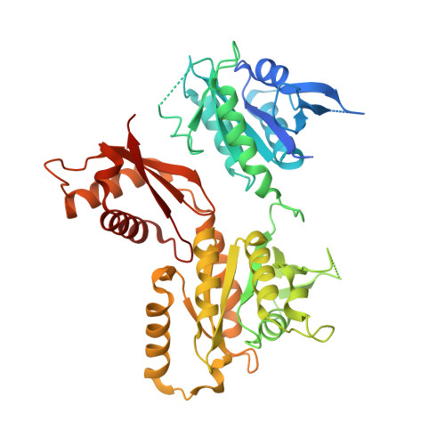
    EngA proteins form a unique family of bacterial GTPases with two GTP-binding domains in tandem, namely GD1 and GD2, followed by a KH (K-homology) domain. They have been shown to interact with the bacterial ribosome and to be involved in its biogenesis. Most prokaryotic EngA possess a high GTPase activity in contrast to eukaryotic GTPases that act mainly as molecular switches. Here, we have purified and characterized the GTPase activity of the Bacillus subtilis EngA and two shortened EngA variants that only contain GD1 or GD2-KH. Interestingly, the GTPase activity of GD1 alone is similar to that of the whole EngA, whereas GD2-KH has a 150-fold lower GTPase activity. At physiological concentration, potassium strongly stimulates the GTPase activity of each protein construct. Interestingly, it affects neither the affinities for nucleotides nor the monomeric status of EngA or the GD1 domain. Thus, potassium likely acts as a chemical GTPase-activating element as proposed for another bacterial GTPase like MnmE. However, unlike MnmE, potassium does not promote dimerization of EngA. In addition, we solved two crystal structures of full-length EngA. One of them contained for the first time a GTP-like analogue bound to GD2 while GD1 was free. Surprisingly, its overall fold was similar to a previously solved structure with GDP bound to both sites. Our data indicate that a significant structural change must occur upon K(+) binding to GD2, and a comparison with T. maritima EngA and MnmE structures allowed us to propose a model explaining the chemical basis for the different GTPase activities of GD1 and GD2.


  • Organizational Affiliation
    • Institut de Biologie Structurale, Université Joseph Fourier Grenoble 1, Grenoble, France.

Macromolecule Content 

  • Total Structure Weight: 51.52 kDa 
  • Atom Count: 3,267 
  • Modeled Residue Count: 402 
  • Deposited Residue Count: 456 
  • Unique protein chains: 1

Macromolecules

Find similar proteins by:|  3D Structure
Entity ID: 1
MoleculeChains  Sequence LengthOrganismDetailsImage
GTP-BINDING PROTEIN ENGA456Bacillus subtilisMutation(s): 0 
Gene Names: derengAyphCBSU22840
UniProt
Find proteins for P50743 (Bacillus subtilis (strain 168))
Explore P50743 
Go to UniProtKB:  P50743
Entity Groups
Sequence Clusters30% Identity50% Identity70% Identity90% Identity95% Identity100% Identity
UniProt GroupP50743
Sequence Annotations
Expand
Reference Sequence

Small Molecules

Ligands 1 Unique
IDChains Name / Formula / InChI Key2D Diagram3D Interactions
GCP

Query on GCP


Download:Ideal Coordinates CCD File
B [auth A]PHOSPHOMETHYLPHOSPHONIC ACID GUANYLATE ESTER
C11 H18 N5 O13 P3
PHBDHXOBFUBCJD-KQYNXXCUSA-N

Experimental Data & Validation

Experimental Data

  • Method: X-RAY DIFFRACTION
  • Resolution: 2.60 Å
  • R-Value Free:  0.268 (Depositor), 0.269 (DCC) 
  • R-Value Work:  0.181 (Depositor), 0.181 (DCC) 
  • R-Value Observed: 0.190 (Depositor) 
Space Group: P 21 21 21
Unit Cell:
Length ( Å )Angle ( ˚ )
a = 62.493α = 90
b = 66.191β = 90
c = 111.74γ = 90
Software Package:
Software NamePurpose
ADSCdata collection
AMoREphasing
REFMACrefinement
XDSdata reduction
XDSdata scaling

Structure Validation

View Full Validation Report


Ligand Structure Quality Assessment 


Entry History 

Deposition Data

Revision History  (Full details and data files)

  • Version 1.0: 2012-11-14
    Type: Initial release
  • Version 1.1: 2023-09-13
    Changes: Data collection, Database references, Derived calculations, Refinement description