4DBZ | pdb_00004dbz

Crystal Structure of V151L Actinorhodin Polyketide Ketoreductase with NADPH


Experimental Data Snapshot

  • Method: X-RAY DIFFRACTION
  • Resolution: 2.64 Å
  • R-Value Free: 
    0.218 (Depositor), 0.223 (DCC) 
  • R-Value Work: 
    0.179 (Depositor), 0.186 (DCC) 
  • R-Value Observed: 
    0.181 (Depositor) 

wwPDB Validation 3D Report Full Report

Validation slider image for 4DBZ

Ligand Structure Quality Assessment 


This is version 1.2 of the entry. See complete history

Literature

The Determinants of Activity and Specificity in Actinorhodin Type II Polyketide Ketoreductase.

Javidpour, P.Bruegger, J.Srithahan, S.Korman, T.P.Crump, M.P.Crosby, J.Burkart, M.D.Tsai, S.C.

(2013) Chem Biol 20: 1225-1234

  • DOI: https://doi.org/10.1016/j.chembiol.2013.07.016
  • Primary Citation Related Structures: 
    4DBZ, 4DC0, 4DC1

  • PubMed Abstract: 

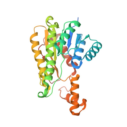
    In the actinorhodin type II polyketide synthase, the first polyketide modification is a regiospecific C9-carbonyl reduction, catalyzed by the ketoreductase (actKR). Our previous studies identified the actKR 94-PGG-96 motif as a determinant of stereospecificity. The molecular basis for reduction regiospecificity is, however, not well understood. In this study, we examined the activities of 20 actKR mutants through a combination of kinetic studies, PKS reconstitution, and structural analyses. Residues have been identified that are necessary for substrate interaction, and these observations have suggested a structural model for this reaction. Polyketides dock at the KR surface and are steered into the enzyme pocket where C7-C12 cyclization is mediated by the KR before C9-ketoreduction can occur. These molecular features can potentially serve as engineering targets for the biosynthesis of novel, reduced polyketides.


  • Organizational Affiliation
    • Department of Molecular Biology and Biochemistry, University of California, Irvine, Irvine, CA 92697, USA.

Macromolecule Content 

  • Total Structure Weight: 60.45 kDa 
  • Atom Count: 3,939 
  • Modeled Residue Count: 518 
  • Deposited Residue Count: 562 
  • Unique protein chains: 1

Macromolecules

Find similar proteins by:|  3D Structure
Entity ID: 1
MoleculeChains  Sequence LengthOrganismDetailsImage
Ketoacyl reductase
A, B
281Streptomyces coelicolorMutation(s): 1 
Gene Names: actIIISCBAC28G1.12cSCO5086
EC: 1.3.1
UniProt
Find proteins for P16544 (Streptomyces coelicolor (strain ATCC BAA-471 / A3(2) / M145))
Explore P16544 
Go to UniProtKB:  P16544
Entity Groups
Sequence Clusters30% Identity50% Identity70% Identity90% Identity95% Identity100% Identity
UniProt GroupP16544
Sequence Annotations
Expand
Reference Sequence

Small Molecules

Ligands 1 Unique
IDChains Name / Formula / InChI Key2D Diagram3D Interactions
NDP

Query on NDP


Download:Ideal Coordinates CCD File
C [auth A],
D [auth B]
NADPH DIHYDRO-NICOTINAMIDE-ADENINE-DINUCLEOTIDE PHOSPHATE
C21 H30 N7 O17 P3
ACFIXJIJDZMPPO-NNYOXOHSSA-N

Experimental Data & Validation

Experimental Data

  • Method: X-RAY DIFFRACTION
  • Resolution: 2.64 Å
  • R-Value Free:  0.218 (Depositor), 0.223 (DCC) 
  • R-Value Work:  0.179 (Depositor), 0.186 (DCC) 
  • R-Value Observed: 0.181 (Depositor) 
Space Group: P 32 2 1
Unit Cell:
Length ( Å )Angle ( ˚ )
a = 102.934α = 90
b = 102.934β = 90
c = 121.844γ = 120
Software Package:
Software NamePurpose
CCP4model building
PHENIXrefinement
HKL-2000data reduction
HKL-2000data scaling
CCP4phasing

Structure Validation

View Full Validation Report


Ligand Structure Quality Assessment 


Entry History 

Deposition Data

Revision History  (Full details and data files)

  • Version 1.0: 2013-01-16
    Type: Initial release
  • Version 1.1: 2013-11-27
    Changes: Database references
  • Version 1.2: 2024-02-28
    Changes: Data collection, Database references, Derived calculations