4DBL | pdb_00004dbl

Crystal structure of E159Q mutant of BtuCDF


Experimental Data Snapshot

  • Method: X-RAY DIFFRACTION
  • Resolution: 3.49 Å
  • R-Value Free: 
    0.251 (Depositor), 0.244 (DCC) 
  • R-Value Work: 
    0.214 (Depositor), 0.207 (DCC) 
  • R-Value Observed: 
    0.215 (Depositor) 

wwPDB Validation 3D Report Full Report

Validation slider image for 4DBL

This is version 1.2 of the entry. See complete history

Literature

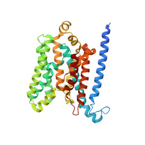
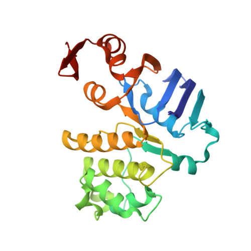
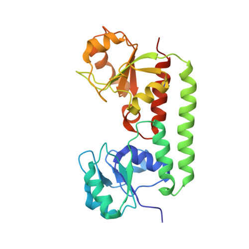
Asymmetric states of vitamin B12 transporter BtuCD are not discriminated by its cognate substrate binding protein BtuF.

Korkhov, V.M.Mireku, S.A.Hvorup, R.N.Locher, K.P.

(2012) FEBS Lett 586: 972-976

  • DOI: https://doi.org/10.1016/j.febslet.2012.02.042
  • Primary Citation Related Structures: 
    4DBL

  • PubMed Abstract: 

    BtuCD is an ABC transporter catalyzing the uptake of vitamin B₁₂ across the Escherichia coli inner membrane. A previously reported X-ray structure of BtuCD in complex with the periplasmic vitamin B₁₂-binding protein BtuF revealed asymmetry of the transmembrane BtuC subunits. The functional relevance of this asymmetry has remained uncertain. Here we report the X-ray structure of a catalytically impaired BtuCD mutant in complex with BtuF, where the BtuC subunits adopt a distinct asymmetric conformation. The structure suggests that BtuF does not discriminate between, or impose, asymmetric conformations of BtuCD. It also explains the conformational disorder observed in BtuCDF crystals.


  • Organizational Affiliation
    • Department of Biology, Institute of Molecular Biology and Biophysics, ETH Zurich, Switzerland.

Macromolecule Content 

  • Total Structure Weight: 317 kDa 
  • Atom Count: 21,230 
  • Modeled Residue Count: 2,778 
  • Deposited Residue Count: 2,902 
  • Unique protein chains: 3

Macromolecules

Find similar proteins by:|  3D Structure
Entity ID: 1
MoleculeChains  Sequence LengthOrganismDetailsImage
Vitamin B12 import system permease protein BtuC
A, B, F, G
349Escherichia coli K-12Mutation(s): 7 
Membrane Entity: Yes 
UniProt
Find proteins for P06609 (Escherichia coli (strain K12))
Explore P06609 
Go to UniProtKB:  P06609
Entity Groups
Sequence Clusters30% Identity50% Identity70% Identity90% Identity95% Identity100% Identity
UniProt GroupP06609
Sequence Annotations
Expand
Reference Sequence
Find similar proteins by:|  3D Structure
Entity ID: 2
MoleculeChains  Sequence LengthOrganismDetailsImage
Vitamin B12 import ATP-binding protein BtuD
C, D, H, I
249Escherichia coli K-12Mutation(s): 2 
EC: 3.6.3.33 (PDB Primary Data), 7.6.2.8 (UniProt)
Membrane Entity: Yes 
UniProt
Find proteins for P06611 (Escherichia coli (strain K12))
Explore P06611 
Go to UniProtKB:  P06611
Entity Groups
Sequence Clusters30% Identity50% Identity70% Identity90% Identity95% Identity100% Identity
UniProt GroupP06611
Sequence Annotations
Expand
Reference Sequence
Find similar proteins by:|  3D Structure
Entity ID: 3
MoleculeChains  Sequence LengthOrganismDetailsImage
Vitamin B12-binding protein
E, J
255Escherichia coli K-12Mutation(s): 0 
Membrane Entity: Yes 
UniProt
Find proteins for P37028 (Escherichia coli (strain K12))
Explore P37028 
Go to UniProtKB:  P37028
Entity Groups
Sequence Clusters30% Identity50% Identity70% Identity90% Identity95% Identity100% Identity
UniProt GroupP37028
Sequence Annotations
Expand
Reference Sequence

Small Molecules

Ligands 2 Unique
IDChains Name / Formula / InChI Key2D Diagram3D Interactions
SO4

Query on SO4


Download:Ideal Coordinates CCD File
L [auth C]
M [auth C]
N [auth C]
P [auth D]
Q [auth D]
L [auth C],
M [auth C],
N [auth C],
P [auth D],
Q [auth D],
R [auth D],
T [auth H],
U [auth H],
V [auth H],
X [auth I],
Y [auth I],
Z [auth I]
SULFATE ION
O4 S
QAOWNCQODCNURD-UHFFFAOYSA-L
PO4

Query on PO4


Download:Ideal Coordinates CCD File
K [auth C],
O [auth D],
S [auth H],
W [auth I]
PHOSPHATE ION
O4 P
NBIIXXVUZAFLBC-UHFFFAOYSA-K

Experimental Data & Validation

Experimental Data

  • Method: X-RAY DIFFRACTION
  • Resolution: 3.49 Å
  • R-Value Free:  0.251 (Depositor), 0.244 (DCC) 
  • R-Value Work:  0.214 (Depositor), 0.207 (DCC) 
  • R-Value Observed: 0.215 (Depositor) 
Space Group: P 1 21 1
Unit Cell:
Length ( Å )Angle ( ˚ )
a = 133.65α = 90
b = 166.93β = 119.76
c = 132.82γ = 90
Software Package:
Software NamePurpose
DENZOdata reduction
PHENIXmodel building
PHENIXrefinement
SCALEPACKdata scaling
PHENIXphasing

Structure Validation

View Full Validation Report


Entry History 

Deposition Data

Revision History  (Full details and data files)

  • Version 1.0: 2012-03-07
    Type: Initial release
  • Version 1.1: 2012-05-23
    Changes: Database references
  • Version 1.2: 2024-10-30
    Changes: Data collection, Database references, Derived calculations, Structure summary