4D7Q | pdb_00004d7q

Crystal structure of a chimeric protein with the Sec7 domain of Legionella pneumophila RalF and the capping domain of Rickettsia prowazekii RalF


Experimental Data Snapshot

  • Method: X-RAY DIFFRACTION
  • Resolution: 2.00 Å
  • R-Value Free: 
    0.217 (Depositor), 0.224 (DCC) 
  • R-Value Work: 
    0.175 (Depositor), 0.183 (DCC) 
  • R-Value Observed: 
    0.177 (Depositor) 

Starting Model: experimental
View more details

wwPDB Validation 3D Report Full Report

Validation slider image for 4D7Q

This is version 1.4 of the entry. See complete history

Literature

On the Use of Legionella/Rickettsia Chimeras to Investigate the Structure and Regulation of Rickettsia Effector Ralf.

Folly-Klan, M.Sancerne, B.Alix, E.Roy, C.R.Cherfils, J.Campanacci, V.

(2015) J Struct Biol 189: 98

  • DOI: https://doi.org/10.1016/j.jsb.2014.12.001
  • Primary Citation Related Structures: 
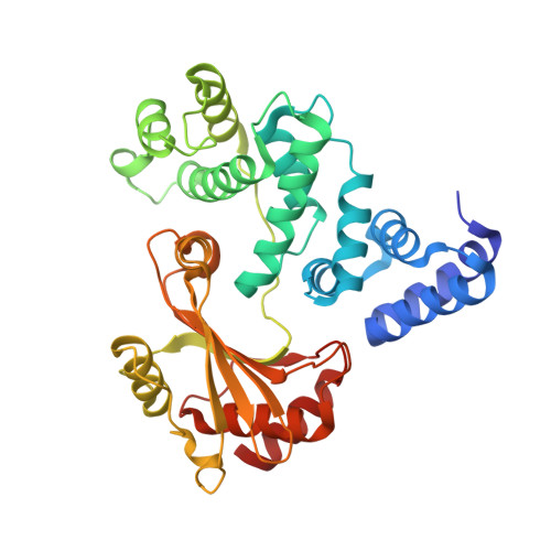
    4D7Q, 4D7R

  • PubMed Abstract: 

    A convenient strategy to interrogate the biology of regulatory proteins is to replace individual domains by an equivalent domain from a related protein of the same species or from an ortholog of another species. It is generally assumed that the overall properties of the native protein are retained in the chimera, and that functional differences reflect only the specific determinants contained in the swapped domains. Here we used this strategy to circumvent the difficulty in obtaining crystals of Rickettsia prowazekii RalF, a bacterial protein that functions as a guanine nucleotide exchange factor for eukaryotic Arf GTPases. A RalF homolog is encoded by Legionella pneumophila, in which a C-terminal capping domain auto-inhibits the catalytic Sec7 domain and localizes the protein to the Legionella-containing vacuole. The crystal structures of domain-swapped chimeras were determined and used to construct a model of Legionella RalF with a RMSD of less than 1Å with the crystal structure, which validated the use of this approach to build a model of Rickettsia RalF. In the Rickettsia RalF model, sequence differences in the capping domain that target it to specific membranes are accommodated by a shift of the entire domain with respect to the Sec7 domain. However, local sequence changes also give rise to an artifactual salt bridge in one of the chimeras, which likely explains why this chimera is recalcitrant to activation. These findings highlight the structural plasticity whereby chimeras can be engineered, but also underline that unpredictable differences can modify their biochemical responses.


  • Organizational Affiliation
    • Laboratoire d'Enzymologie et Biochimie Structurales, CNRS, Centre de Recherche de Gif, 91198 Gif-sur-Yvette, France.

Macromolecule Content 

  • Total Structure Weight: 41.4 kDa 
  • Atom Count: 3,145 
  • Modeled Residue Count: 347 
  • Deposited Residue Count: 352 
  • Unique protein chains: 1

Macromolecules

Find similar proteins by:|  3D Structure
Entity ID: 1
MoleculeChains  Sequence LengthOrganismDetailsImage
RALF, PROLINE/BETAINE TRANSPORTER352Legionella pneumophila subsp. pneumophila str. Philadelphia 1Rickettsia prowazekii str. Madrid E
This entity is chimeric
Mutation(s): 2 
UniProt
Find proteins for Q8RT31 (Legionella pneumophila)
Explore Q8RT31 
Go to UniProtKB:  Q8RT31
Find proteins for Q9ZDF5 (Rickettsia prowazekii (strain Madrid E))
Explore Q9ZDF5 
Go to UniProtKB:  Q9ZDF5
Entity Groups
Sequence Clusters30% Identity50% Identity70% Identity90% Identity95% Identity100% Identity
UniProt GroupsQ8RT31Q9ZDF5
Sequence Annotations
Expand
Reference Sequence

Small Molecules

Ligands 4 Unique
IDChains Name / Formula / InChI Key2D Diagram3D Interactions
EPE

Query on EPE


Download:Ideal Coordinates CCD File
B [auth A]4-(2-HYDROXYETHYL)-1-PIPERAZINE ETHANESULFONIC ACID
C8 H18 N2 O4 S
JKMHFZQWWAIEOD-UHFFFAOYSA-N
PEG

Query on PEG


Download:Ideal Coordinates CCD File
H [auth A],
I [auth A]
DI(HYDROXYETHYL)ETHER
C4 H10 O3
MTHSVFCYNBDYFN-UHFFFAOYSA-N
GOL

Query on GOL


Download:Ideal Coordinates CCD File
D [auth A],
E [auth A],
F [auth A],
G [auth A]
GLYCEROL
C3 H8 O3
PEDCQBHIVMGVHV-UHFFFAOYSA-N
NO3

Query on NO3


Download:Ideal Coordinates CCD File
C [auth A]NITRATE ION
N O3
NHNBFGGVMKEFGY-UHFFFAOYSA-N

Experimental Data & Validation

Experimental Data

  • Method: X-RAY DIFFRACTION
  • Resolution: 2.00 Å
  • R-Value Free:  0.217 (Depositor), 0.224 (DCC) 
  • R-Value Work:  0.175 (Depositor), 0.183 (DCC) 
  • R-Value Observed: 0.177 (Depositor) 
Space Group: P 21 21 21
Unit Cell:
Length ( Å )Angle ( ˚ )
a = 40.31α = 90
b = 77.44β = 90
c = 119.11γ = 90
Software Package:
Software NamePurpose
BUSTERrefinement
XDSdata reduction
XDSdata scaling
PHASERphasing

Structure Validation

View Full Validation Report


Entry History 

Deposition Data

Revision History  (Full details and data files)

  • Version 1.0: 2015-01-14
    Type: Initial release
  • Version 1.1: 2015-01-21
    Changes: Atomic model, Derived calculations, Other
  • Version 1.2: 2015-02-18
    Changes: Database references
  • Version 1.3: 2017-03-15
    Changes: Source and taxonomy
  • Version 1.4: 2023-12-20
    Changes: Data collection, Database references, Derived calculations, Other, Refinement description