


From Mm Fragments to Nm Compounds Using Iloe-NMR to Guide Linking of Compounds in Fragment Based Drug Discovery (Fbdd).
Jacso, T., Ullah, V., Sandmark, J., Oster, L., Redzick, A., Borjesson, U., Olsson, T., Akerud, T.To be published.
Experimental Data Snapshot
Entity ID: 1 | |||||
|---|---|---|---|---|---|
| Molecule | Chains | Sequence Length | Organism | Details | Image |
| COAGULATION FACTOR XI | 238 | Homo sapiens | Mutation(s): 4 EC: 3.4.21.27 | 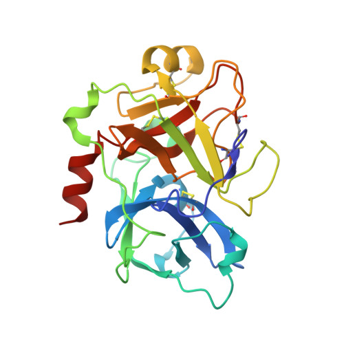 | |
UniProt & NIH Common Fund Data Resources | |||||
PHAROS: P03951 GTEx: ENSG00000088926 | |||||
Entity Groups | |||||
| Sequence Clusters | 30% Identity50% Identity70% Identity90% Identity95% Identity100% Identity | ||||
| UniProt Group | P03951 | ||||
Sequence AnnotationsExpand | |||||
Reference Sequence | |||||
| Ligands 3 Unique | |||||
|---|---|---|---|---|---|
| ID | Chains | Name / Formula / InChI Key | 2D Diagram | 3D Interactions | |
| IXA Download:Ideal Coordinates CCD File | C [auth A] | N-[(2S)-1-({4-[(diaminomethylidene)amino]butyl}amino)-1-oxo-3-phenylpropan-2-yl]-4-hydroxy-2-oxo-1,2-dihydroquinoline-6-carboxamide C24 H28 N6 O4 DZWHLWITPPCGRM-IBGZPJMESA-N |  | ||
| SO4 Download:Ideal Coordinates CCD File | D [auth A], E [auth A], F [auth A], G [auth A] | SULFATE ION O4 S QAOWNCQODCNURD-UHFFFAOYSA-L |  | ||
| GOL Download:Ideal Coordinates CCD File | B [auth A] | GLYCEROL C3 H8 O3 PEDCQBHIVMGVHV-UHFFFAOYSA-N |  | ||
| Length ( Å ) | Angle ( ˚ ) |
|---|---|
| a = 121.293 | α = 90 |
| b = 121.293 | β = 90 |
| c = 121.293 | γ = 90 |
| Software Name | Purpose |
|---|---|
| REFMAC | refinement |
| MOSFLM | data reduction |
| Aimless | data scaling |
| PHASER | phasing |


