4D7C

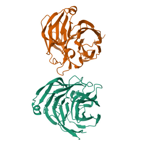
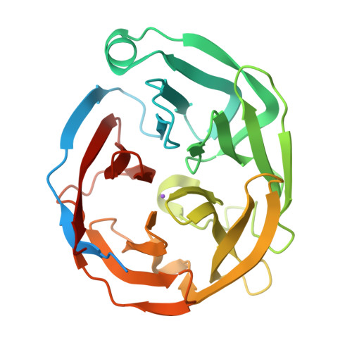
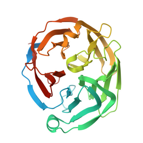
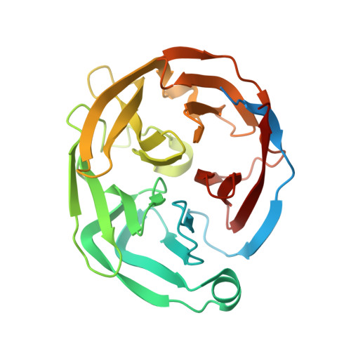
Monoclinic crystal form of the extracellular olfactomedin domain from gliomedin


Experimental Data Snapshot

  • Method: X-RAY DIFFRACTION
  • Resolution: 1.45 Å
  • R-Value Free: 0.212 
  • R-Value Work: 0.173 
  • R-Value Observed: 0.174 

Starting Model: experimental
View more details

wwPDB Validation   3D Report Full Report


This is version 1.3 of the entry. See complete history


Literature

The Olfactomedin Domain from Gliomedin is a Beta-Propeller with Unique Structural Properties.

Han, H.Kursula, P.

(2015) J Biol Chem 290: 3612

  • DOI: https://doi.org/10.1074/jbc.M114.627547
  • Primary Citation of Related Structures:  
    4D77, 4D7C

  • PubMed Abstract: 

    All members of the olfactomedin (OLF) family have a conserved extracellular OLF domain, for which a structure has not been available. We present here the crystal structure of the OLF domain from gliomedin. Gliomedin is a protein expressed by Schwann cells in peripheral nerves, important for the formation of the nodes of Ranvier. Gliomedin interacts with neuronal cell adhesion molecules, such as neurofascin, but the structural details of the interaction are not known. The structure of the OLF domain presents a five-bladed β-propeller fold with unusual geometric properties. The symmetry of the structure is not 5-fold, but rather reveals a twisted arrangement. The conserved top face of the gliomedin OLF domain is likely to be important for binding to neuronal ligands. Our results provide a structural basis for the functions of gliomedin in Schwann cells, enable the understanding of the role of the gliomedin OLF domain in autoimmune neuropathies, and unravel the locations of human disease-causing mutations in other OLF family members, including myocilin.


  • Organizational Affiliation

    From the Faculty of Biochemistry and Molecular Medicine and Biocenter Oulu, University of Oulu, 90014 Oulu, Finland, the German Electron Synchrotron (DESY), 22607 Hamburg, Germany, and.


Macromolecules
Find similar proteins by:  (by identity cutoff)  |  3D Structure
Entity ID: 1
MoleculeChains Sequence LengthOrganismDetailsImage
GLIOMEDIN
A, B
285Rattus norvegicusMutation(s): 0 
UniProt
Find proteins for Q80WL1 (Rattus norvegicus)
Explore Q80WL1 
Go to UniProtKB:  Q80WL1
Entity Groups  
Sequence Clusters30% Identity50% Identity70% Identity90% Identity95% Identity100% Identity
UniProt GroupQ80WL1
Sequence Annotations
Expand
  • Reference Sequence
Experimental Data & Validation

Experimental Data

  • Method: X-RAY DIFFRACTION
  • Resolution: 1.45 Å
  • R-Value Free: 0.212 
  • R-Value Work: 0.173 
  • R-Value Observed: 0.174 
  • Space Group: P 1 21 1
Unit Cell:
Length ( Å )Angle ( ˚ )
a = 37.55α = 90
b = 141.7β = 110.61
c = 46.03γ = 90
Software Package:
Software NamePurpose
PHENIXrefinement
XDSdata reduction
XDSdata scaling
PHASERphasing

Structure Validation

View Full Validation Report


Entry History 

Deposition Data

Revision History  (Full details and data files)

  • Version 1.0: 2014-12-24
    Type: Initial release
  • Version 1.1: 2014-12-31
    Changes: Database references
  • Version 1.2: 2015-03-04
    Changes: Database references
  • Version 1.3: 2023-12-20
    Changes: Data collection, Database references, Derived calculations, Other, Refinement description