4D69 | pdb_00004d69

SOYBEAN AGGLUTININ FROM GLYCINE MAX IN COMPLEX WITH THE ANTIGEN Tn


Experimental Data Snapshot

  • Method: X-RAY DIFFRACTION
  • Resolution: 2.70 Å
  • R-Value Free: 
    0.274 (Depositor), 0.271 (DCC) 
  • R-Value Work: 
    0.230 (Depositor), 0.233 (DCC) 
  • R-Value Observed: 
    0.231 (Depositor) 

Starting Model: experimental
View more details

wwPDB Validation 3D Report Full Report

Validation slider image for 4D69

Ligand Structure Quality Assessment 


This is version 2.2 of the entry. See complete history

Literature

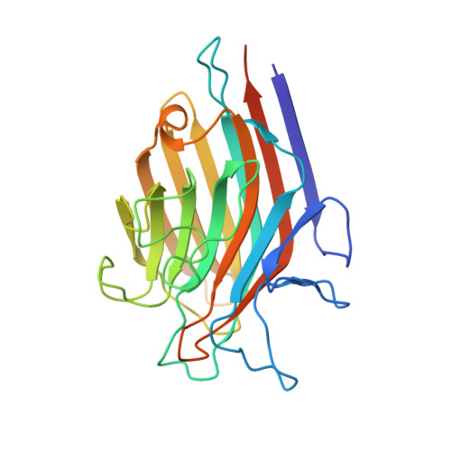
Detection of Tumor-Associated Glycopeptides by Lectins: The Peptide Context Modulates Carbohydrate Recognition.

Madariaga, D.Martinez-Saez, N.Somovilla, V.J.Coelho, H.Valero-Gonzalez, J.Castro-Lopez, J.Asensio, J.L.Jimenez-Barbero, J.Busto, J.H.Avenoza, A.Marcelo, F.Hurtado-Guerrero, R.Corzana, F.Peregrina, J.M.

(2015) ACS Chem Biol 10: 747

  • DOI: https://doi.org/10.1021/cb500855x
  • Primary Citation Related Structures: 
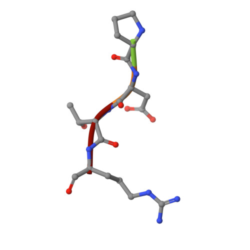
    4D69

  • PubMed Abstract: 

    Tn antigen (α-O-GalNAc-Ser/Thr) is a convenient cancer biomarker that is recognized by antibodies and lectins. This work yields remarkable results for two plant lectins in terms of epitope recognition and reveals that these receptors show higher affinity for Tn antigen when it is incorporated in the Pro-Asp-Thr-Arg (PDTR) peptide region of mucin MUC1. In contrast, a significant affinity loss is observed when Tn antigen is located in the Ala-His-Gly-Val-Thr-Ser-Ala (AHGVTSA) or Ala-Pro-Gly-Ser-Thr-Ala-Pro (APGSTAP) fragments. Our data indicate that the charged residues, Arg and Asp, present in the PDTR sequence establish noteworthy fundamental interactions with the lectin surface as well as fix the conformation of the peptide backbone, favoring the presentation of the sugar moiety toward the lectin. These results may help to better understand glycopeptide-lectin interactions and may contribute to engineer new binding sites, allowing novel glycosensors for Tn antigen detection to be designed.


  • Organizational Affiliation
    • Centro de Investigación en Síntesis Química, Departamento de Química, Universidad de La Rioja, E-26006 Logroño, Spain.

Macromolecule Content 

  • Total Structure Weight: 346.93 kDa 
  • Atom Count: 22,512 
  • Modeled Residue Count: 2,822 
  • Deposited Residue Count: 3,096 
  • Unique protein chains: 2

Macromolecules

Find similar proteins by:|  3D Structure
Entity ID: 1
MoleculeChains  Sequence LengthOrganismDetailsImage
LECTIN
A, B, C, D, E
A, B, C, D, E, F, G, H, I, J, K, L
253Glycine maxMutation(s): 0 
UniProt
Find proteins for P05046 (Glycine max)
Explore P05046 
Go to UniProtKB:  P05046
Entity Groups
Sequence Clusters30% Identity50% Identity70% Identity90% Identity95% Identity100% Identity
UniProt GroupP05046
Glycosylation
Glycosylation Sites: 1
Sequence Annotations
Expand
Reference Sequence
Find similar proteins by:  Sequence   |   3D Structure  

Oligosaccharides

Help  
Entity ID: 3
MoleculeChains Length2D Diagram GlycosylationD Interactions
2-acetamido-2-deoxy-beta-D-glucopyranose-(1-4)-2-acetamido-2-deoxy-beta-D-glucopyranose
AA [auth a],
BA [auth b],
CA [auth c],
DA [auth d],
EA [auth e],
AA [auth a],
BA [auth b],
CA [auth c],
DA [auth d],
EA [auth e],
FA [auth f],
GA [auth g],
HA [auth h],
IA [auth i],
JA [auth j],
Y [auth M],
Z [auth N]
2N-Glycosylation
Glycosylation Resources
GlyTouCan: G42666HT
GlyCosmos: G42666HT
GlyGen: G42666HT

Small Molecules

Ligands 2 Unique
IDChains Name / Formula / InChI Key2D Diagram3D Interactions
A2G

Query on A2G


Download:Ideal Coordinates CCD File
BB [auth F]
CB [auth G]
FB [auth H]
IB [auth I]
KA [auth A]
BB [auth F],
CB [auth G],
FB [auth H],
IB [auth I],
KA [auth A],
LB [auth J],
NA [auth B],
OB [auth K],
QA [auth C],
RB [auth L],
TA [auth D],
WA [auth E]
2-acetamido-2-deoxy-alpha-D-galactopyranose
C8 H15 N O6
OVRNDRQMDRJTHS-CBQIKETKSA-N
MN

Query on MN


Download:Ideal Coordinates CCD File
AB [auth F]
DB [auth G]
EB [auth G]
GB [auth H]
HB [auth H]
AB [auth F],
DB [auth G],
EB [auth G],
GB [auth H],
HB [auth H],
JB [auth I],
KB [auth I],
LA [auth A],
MA [auth A],
MB [auth J],
NB [auth J],
OA [auth B],
PA [auth B],
PB [auth K],
QB [auth K],
RA [auth C],
SA [auth C],
SB [auth L],
TB [auth L],
UA [auth D],
VA [auth D],
XA [auth E],
YA [auth E],
ZA [auth F]
MANGANESE (II) ION
Mn
WAEMQWOKJMHJLA-UHFFFAOYSA-N

Experimental Data & Validation

Experimental Data

  • Method: X-RAY DIFFRACTION
  • Resolution: 2.70 Å
  • R-Value Free:  0.274 (Depositor), 0.271 (DCC) 
  • R-Value Work:  0.230 (Depositor), 0.233 (DCC) 
  • R-Value Observed: 0.231 (Depositor) 
Space Group: P 32
Unit Cell:
Length ( Å )Angle ( ˚ )
a = 114.232α = 90
b = 114.232β = 90
c = 202.893γ = 120
Software Package:
Software NamePurpose
REFMACrefinement
SCALEPACKdata scaling
PHASERphasing

Structure Validation

View Full Validation Report


Ligand Structure Quality Assessment 


Entry History 

Revision History  (Full details and data files)

  • Version 1.0: 2014-11-19
    Type: Initial release
  • Version 1.1: 2014-12-17
    Changes: Database references
  • Version 1.2: 2015-04-15
    Changes: Database references
  • Version 2.0: 2020-07-29
    Type: Remediation
    Reason: Carbohydrate remediation
    Changes: Advisory, Atomic model, Data collection, Derived calculations, Other, Structure summary
  • Version 2.1: 2023-12-20
    Changes: Data collection, Database references, Refinement description, Structure summary
  • Version 2.2: 2024-11-13
    Changes: Structure summary