4D4X | pdb_00004d4x

Nitrosyl complex of the D121I variant of cytochrome c prime from Alcaligenes xylosoxidans


Experimental Data Snapshot

  • Method: X-RAY DIFFRACTION
  • Resolution: 1.30 Å
  • R-Value Free: 
    0.214 (Depositor), 0.217 (DCC) 
  • R-Value Work: 
    0.169 (Depositor), 0.179 (DCC) 
  • R-Value Observed: 
    0.171 (Depositor) 

wwPDB Validation 3D Report Full Report

Validation slider image for 4D4X

Ligand Structure Quality Assessment 


This is version 2.1 of the entry. See complete history

Literature

Hydrogen Bonding of the Dissociated Histidine Ligand is not Required for Formation of a Proximal No Adduct in Cytochrome C'.

Ghafoor, D.D.Kekilli, D.Abdullah, G.H.Dworkowski, F.S.N.Hassan, H.G.Wilson, M.T.Strange, R.W.Hough, M.A.

(2015) J Biol Inorg Chem 20: 949

  • DOI: https://doi.org/10.1007/s00775-015-1278-y
  • Primary Citation Related Structures: 
    4D4N, 4D4X, 5AGF

  • PubMed Abstract: 

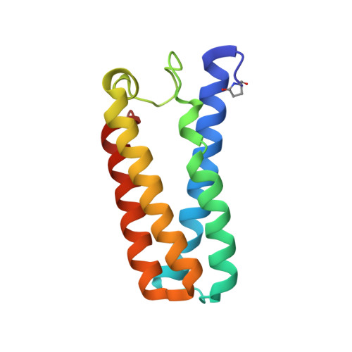
    Cytochromes c', that occur in methanotrophic, denitrifying and photosynthetic bacteria, form unusual proximal penta-coordinate NO complexes via a hexa-coordinate distal NO intermediate. Their NO binding properties are similar to those of the eukaryotic NO sensor, soluble guanylate cyclase, for which they provide a valuable structural model. Previous studies suggested that hydrogen bonding between the displaced proximal histidine (His120) ligand (following its dissociation from heme due to trans effects from the distally bound NO) and a conserved aspartate residue (Asp121) could play a key role in allowing proximal NO binding to occur. We have characterized three variants of Alcaligenes xylosoxidans cytochrome c' (AXCP) where Asp121 has been replaced by Ala, Ile and Gln, respectively. In all variants, hydrogen bonding between residue 121 and His120 is abolished yet 5-coordinate proximal NO species are still formed. Our data therefore demonstrate that the His120-Asp121 bond is not essential for proximal NO binding although it likely provides an energy minimum for the displaced His ligand. All variants have altered proximal pocket structure relative to native AXCP.


  • Organizational Affiliation
    • Faculty of Science and Education Science, University of Sulaimani, Sulaymaniyah, Iraq.

Macromolecule Content 

  • Total Structure Weight: 14.28 kDa 
  • Atom Count: 1,214 
  • Modeled Residue Count: 126 
  • Deposited Residue Count: 127 
  • Unique protein chains: 1

Macromolecules

Find similar proteins by:|  3D Structure
Entity ID: 1
MoleculeChains  Sequence LengthOrganismDetailsImage
CYTOCHROME C'127Achromobacter xylosoxidansMutation(s): 1 
UniProt
Find proteins for P00138 (Alcaligenes xylosoxydans xylosoxydans)
Explore P00138 
Go to UniProtKB:  P00138
Entity Groups
Sequence Clusters30% Identity50% Identity70% Identity90% Identity95% Identity100% Identity
UniProt GroupP00138
Sequence Annotations
Expand
Reference Sequence

Small Molecules

Modified Residues  1 Unique
IDChains TypeFormula2D DiagramParent
PCA
Query on PCA
A
L-PEPTIDE LINKINGC5 H7 N O3GLN

Experimental Data & Validation

Experimental Data

  • Method: X-RAY DIFFRACTION
  • Resolution: 1.30 Å
  • R-Value Free:  0.214 (Depositor), 0.217 (DCC) 
  • R-Value Work:  0.169 (Depositor), 0.179 (DCC) 
  • R-Value Observed: 0.171 (Depositor) 
Space Group: P 65 2 2
Unit Cell:
Length ( Å )Angle ( ˚ )
a = 53.155α = 90
b = 53.155β = 90
c = 180.501γ = 120
Software Package:
Software NamePurpose
REFMACrefinement
XDSdata reduction
Aimlessdata scaling

Structure Validation

View Full Validation Report


Ligand Structure Quality Assessment 


Entry History 

Deposition Data

Revision History  (Full details and data files)

  • Version 1.0: 2015-09-09
    Type: Initial release
  • Version 2.0: 2020-03-11
    Changes: Advisory, Derived calculations, Other, Polymer sequence
  • Version 2.1: 2024-11-13
    Changes: Advisory, Data collection, Database references, Derived calculations, Structure summary