


Pyridazinones: A Novel Scaffold with Excellent Physicochemical Properties and Safety Profile for a Clinically Validated Target of Mycobacterium Tuberculosis
Lange, S.To be published.
Experimental Data Snapshot
Starting Model: experimental
View more details
Entity ID: 1 | |||||
|---|---|---|---|---|---|
| Molecule | Chains | Sequence Length | Organism | Details | Image |
| ENOYL-[ACYL-CARRIER-PROTEIN] REDUCTASE [NADH] | 269 | Mycobacterium tuberculosis H37Rv | Mutation(s): 0 EC: 1.3.1.9 | 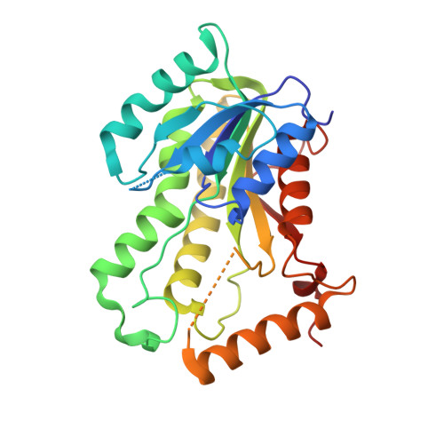 | |
UniProt | |||||
Entity Groups | |||||
| Sequence Clusters | 30% Identity50% Identity70% Identity90% Identity95% Identity100% Identity | ||||
| UniProt Group | P9WGR1 | ||||
Sequence AnnotationsExpand | |||||
Reference Sequence | |||||
| Ligands 3 Unique | |||||
|---|---|---|---|---|---|
| ID | Chains | Name / Formula / InChI Key | 2D Diagram | 3D Interactions | |
| NAD Download:Ideal Coordinates CCD File | E [auth A], H [auth B], J [auth C], L [auth D] | NICOTINAMIDE-ADENINE-DINUCLEOTIDE C21 H27 N7 O14 P2 BAWFJGJZGIEFAR-NNYOXOHSSA-N |  | ||
| 9G4 Download:Ideal Coordinates CCD File | F [auth A], I [auth B], K [auth C], M [auth D] | 1-{4-[(acetylamino)methyl]phenyl}-4-(4-chlorophenoxy)-6-oxo-1,6-dihydropyridazine-3-carboxamide C20 H17 Cl N4 O4 SYAJIRKWQQECEF-UHFFFAOYSA-N |  | ||
| MG Download:Ideal Coordinates CCD File | G [auth A], N [auth D] | MAGNESIUM ION Mg JLVVSXFLKOJNIY-UHFFFAOYSA-N |  | ||
| Length ( Å ) | Angle ( ˚ ) |
|---|---|
| a = 64.916 | α = 90 |
| b = 111.421 | β = 98.46 |
| c = 67.663 | γ = 90 |
| Software Name | Purpose |
|---|---|
| BUSTER | refinement |
| XDS | data reduction |
| Aimless | data scaling |
| AMoRE | phasing |


