4D04 | pdb_00004d04

Structure of the Cys65Asp mutant of phenylacetone monooxygenase: reduced state


Experimental Data Snapshot

  • Method: X-RAY DIFFRACTION
  • Resolution: 1.75 Å
  • R-Value Free: 
    0.216 (Depositor), 0.219 (DCC) 
  • R-Value Work: 
    0.183 (Depositor), 0.190 (DCC) 
  • R-Value Observed: 
    0.184 (Depositor) 

Starting Model: experimental
View more details

wwPDB Validation 3D Report Full Report

Validation slider image for 4D04

Ligand Structure Quality Assessment 


This is version 1.2 of the entry. See complete history

Literature

Finding the Switch: Turning a Baeyer-Villiger Monooxygenase Into a Nadph Oxidase.

Brondani, P.B.Dudek, H.M.Martinoli, C.Mattevi, A.Fraaije, M.W.

(2014) J Am Chem Soc 136: 16966

  • DOI: https://doi.org/10.1021/ja508265b
  • Primary Citation Related Structures: 
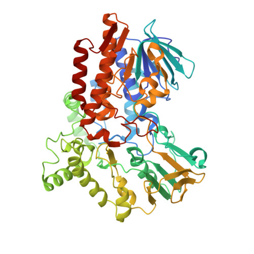
    4D03, 4D04

  • PubMed Abstract: 

    By a targeted enzyme engineering approach, we were able to create an efficient NADPH oxidase from a monooxygenase. Intriguingly, replacement of only one specific single amino acid was sufficient for such a monooxygenase-to-oxidase switch-a complete transition in enzyme activity. Pre-steady-state kinetic analysis and elucidation of the crystal structure of the C65D PAMO mutant revealed that the mutation introduces small changes near the flavin cofactor, resulting in a rapid decay of the peroxyflavin intermediate. The engineered biocatalyst was shown to be a thermostable, solvent tolerant, and effective cofactor-regenerating biocatalyst. Therefore, it represents a valuable new biocatalytic tool.


  • Organizational Affiliation
    • Molecular Enzymology Group, University of Groningen , Nijenborgh 4, 9747AG Groningen, The Netherlands.

Macromolecule Content 

  • Total Structure Weight: 63.3 kDa 
  • Atom Count: 4,678 
  • Modeled Residue Count: 532 
  • Deposited Residue Count: 542 
  • Unique protein chains: 1

Macromolecules

Find similar proteins by:|  3D Structure
Entity ID: 1
MoleculeChains  Sequence LengthOrganismDetailsImage
PHENYLACETONE MONOOXYGENASE542Thermobifida fuscaMutation(s): 1 
EC: 1.14.13.92
UniProt
Find proteins for Q47PU3 (Thermobifida fusca (strain YX))
Explore Q47PU3 
Go to UniProtKB:  Q47PU3
Entity Groups
Sequence Clusters30% Identity50% Identity70% Identity90% Identity95% Identity100% Identity
UniProt GroupQ47PU3
Sequence Annotations
Expand
Reference Sequence

Experimental Data & Validation

Experimental Data

  • Method: X-RAY DIFFRACTION
  • Resolution: 1.75 Å
  • R-Value Free:  0.216 (Depositor), 0.219 (DCC) 
  • R-Value Work:  0.183 (Depositor), 0.190 (DCC) 
  • R-Value Observed: 0.184 (Depositor) 
Space Group: P 32 2 1
Unit Cell:
Length ( Å )Angle ( ˚ )
a = 107.879α = 90
b = 107.879β = 90
c = 107.044γ = 120
Software Package:
Software NamePurpose
REFMACrefinement
MOSFLMdata reduction
Aimlessdata scaling
PHASERphasing

Structure Validation

View Full Validation Report


Ligand Structure Quality Assessment 


Entry History 

Deposition Data

Revision History  (Full details and data files)

  • Version 1.0: 2014-12-10
    Type: Initial release
  • Version 1.1: 2014-12-24
    Changes: Database references
  • Version 1.2: 2023-12-20
    Changes: Data collection, Database references, Derived calculations, Other, Refinement description