4CZB | pdb_00004czb

Structure of the sodium proton antiporter MjNhaP1 from Methanocaldococcus jannaschii at pH 8.


Experimental Data Snapshot

  • Method: X-RAY DIFFRACTION
  • Resolution: 3.50 Å
  • R-Value Free: 
    0.302 (Depositor), 0.305 (DCC) 
  • R-Value Work: 
    0.252 (Depositor), 0.261 (DCC) 

Starting Model: experimental
View more details

wwPDB Validation 3D Report Full Report

Validation slider image for 4CZB

This is version 1.1 of the entry. See complete history

Literature

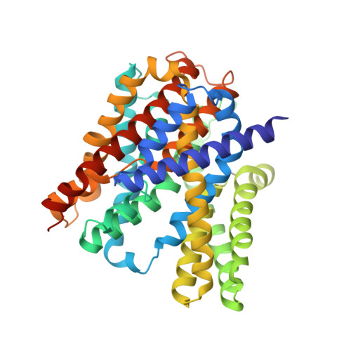
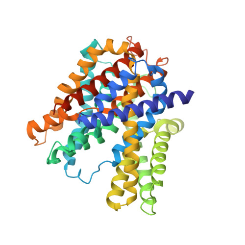
3D Em Map of the Sodium Proton Antiporter Mjnhap1 from Methanocaldococcus Jannaschii

Paulino, C.Woehlert, D.Yildiz, O.Kuhlbrandt, W.

(2014) Elife 3

  • DOI: https://doi.org/10.7554/eLife.03583
  • Primary Citation Related Structures: 
    4CZB, 4D0A

  • PubMed Abstract: 

    Sodium/proton antiporters are essential for sodium and pH homeostasis and play a major role in human health and disease. We determined the structures of the archaeal sodium/proton antiporter MjNhaP1 in two complementary states. The inward-open state was obtained by x-ray crystallography in the presence of sodium at pH 8, where the transporter is highly active. The outward-open state was obtained by electron crystallography without sodium at pH 4, where MjNhaP1 is inactive. Comparison of both structures reveals a 7° tilt of the 6 helix bundle. (22)Na(+) uptake measurements indicate non-cooperative transport with an activity maximum at pH 7.5. We conclude that binding of a Na(+) ion from the outside induces helix movements that close the extracellular cavity, open the cytoplasmic funnel, and result in a ∼5 Å vertical relocation of the ion binding site to release the substrate ion into the cytoplasm.


  • Organizational Affiliation
    • Department of Structural Biology, Max Planck Institute of Biophysics, Frankfurt am Main, Germany.

Macromolecule Content 

  • Total Structure Weight: 184.25 kDa 
  • Atom Count: 12,548 
  • Modeled Residue Count: 1,655 
  • Deposited Residue Count: 1,704 
  • Unique protein chains: 2

Macromolecules

Find similar proteins by:|  3D Structure
Entity ID: 1
MoleculeChains  Sequence LengthOrganismDetailsImage
NA(+)/H(+) ANTIPORTER 1
A, B, D
426Methanocaldococcus jannaschiiMutation(s): 0 
Membrane Entity: Yes 
UniProt
Find proteins for Q60362 (Methanocaldococcus jannaschii (strain ATCC 43067 / DSM 2661 / JAL-1 / JCM 10045 / NBRC 100440))
Explore Q60362 
Go to UniProtKB:  Q60362
Entity Groups
Sequence Clusters30% Identity50% Identity70% Identity90% Identity95% Identity100% Identity
UniProt GroupQ60362
Sequence Annotations
Expand
Reference Sequence
Find similar proteins by:|  3D Structure
Entity ID: 2
MoleculeChains  Sequence LengthOrganismDetailsImage
NA(+)/H(+) ANTIPORTER 1426Methanocaldococcus jannaschiiMutation(s): 0 
Membrane Entity: Yes 
UniProt
Find proteins for Q60362 (Methanocaldococcus jannaschii (strain ATCC 43067 / DSM 2661 / JAL-1 / JCM 10045 / NBRC 100440))
Explore Q60362 
Go to UniProtKB:  Q60362
Entity Groups
Sequence Clusters30% Identity50% Identity70% Identity90% Identity95% Identity100% Identity
UniProt GroupQ60362
Sequence Annotations
Expand
Reference Sequence

Experimental Data & Validation

Experimental Data

  • Method: X-RAY DIFFRACTION
  • Resolution: 3.50 Å
  • R-Value Free:  0.302 (Depositor), 0.305 (DCC) 
  • R-Value Work:  0.252 (Depositor), 0.261 (DCC) 
Space Group: P 1 21 1
Unit Cell:
Length ( Å )Angle ( ˚ )
a = 98.437α = 90
b = 102.544β = 105.63
c = 132.096γ = 90
Software Package:
Software NamePurpose
PHENIXrefinement
PHASERphasing

Structure Validation

View Full Validation Report


Entry History 

Deposition Data

Revision History  (Full details and data files)

  • Version 1.0: 2014-12-10
    Type: Initial release
  • Version 1.1: 2023-12-20
    Changes: Data collection, Database references, Derived calculations, Other, Refinement description