4CYG | pdb_00004cyg

The structure of vanin-1: defining the link between metabolic disease, oxidative stress and inflammation


Experimental Data Snapshot

  • Method: X-RAY DIFFRACTION
  • Resolution: 2.30 Å
  • R-Value Free: 
    0.214 (Depositor), 0.217 (DCC) 
  • R-Value Work: 
    0.186 (Depositor), 0.193 (DCC) 
  • R-Value Observed: 
    0.188 (Depositor) 

Starting Model: experimental
View more details

wwPDB Validation 3D Report Full Report

Validation slider image for 4CYG

Ligand Structure Quality Assessment 


This is version 2.2 of the entry. See complete history

Literature

The Structure of Vanin-1: A Key Enzyme Linking Metabolic Disease and Inflammation

Boersma, Y.L.Newman, J.Adams, T.E.Cowieson, N.Krippner, G.Bozaoglu, K.Peat, T.S.

(2014) Acta Crystallogr D Biol Crystallogr 70: 3320

  • DOI: https://doi.org/10.1107/S1399004714022767
  • Primary Citation Related Structures: 
    4CYF, 4CYG, 4CYY

  • PubMed Abstract: 

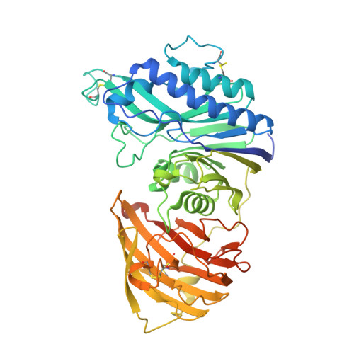
    Although part of the coenzyme A pathway, vanin 1 (also known as pantetheinase) sits on the cell surface of many cell types as an ectoenzyme, catalyzing the breakdown of pantetheine to pantothenic acid (vitamin B5) and cysteamine, a strong reducing agent. Vanin 1 was initially discovered as a protein involved in the homing of leukocytes to the thymus. Numerous studies have shown that vanin 1 is involved in inflammation, and more recent studies have shown a key role in metabolic disease. Here, the X-ray crystal structure of human vanin 1 at 2.25 Å resolution is presented, which is the first reported structure from the vanin family, as well as a crystal structure of vanin 1 bound to a specific inhibitor. These structures illuminate how vanin 1 can mediate its biological roles by way of both enzymatic activity and protein-protein interactions. Furthermore, it sheds light on how the enzymatic activity is regulated by a novel allosteric mechanism at a domain interface.


  • Organizational Affiliation
    • Department of Pharmaceutical Biology, Groningen Research Institute of Pharmacy, University of Groningen, A. Deusinglaan 1, 9713 AV Groningen, The Netherlands.

Macromolecule Content 

  • Total Structure Weight: 115.21 kDa 
  • Atom Count: 7,820 
  • Modeled Residue Count: 925 
  • Deposited Residue Count: 1,012 
  • Unique protein chains: 1

Macromolecules

Find similar proteins by:|  3D Structure
Entity ID: 1
MoleculeChains  Sequence LengthOrganismDetailsImage
PANTETHEINASE
A, B
506Homo sapiensMutation(s): 1 
EC: 3.5.1.92
UniProt & NIH Common Fund Data Resources
Find proteins for O95497 (Homo sapiens)
Explore O95497 
Go to UniProtKB:  O95497
PHAROS:  O95497
GTEx:  ENSG00000112299 
Entity Groups
Sequence Clusters30% Identity50% Identity70% Identity90% Identity95% Identity100% Identity
UniProt GroupO95497
Glycosylation
Glycosylation Sites: 4Go to GlyGen: O95497-1
Sequence Annotations
Expand
Reference Sequence

Oligosaccharides

Help  
Entity ID: 2
MoleculeChains Length2D Diagram GlycosylationD Interactions
2-acetamido-2-deoxy-beta-D-glucopyranose-(1-4)-2-acetamido-2-deoxy-beta-D-glucopyranose
C
2N-Glycosylation
Glycosylation Resources
GlyTouCan: G42666HT
GlyCosmos: G42666HT
GlyGen: G42666HT

Small Molecules

Ligands 3 Unique
IDChains Name / Formula / InChI Key2D Diagram3D Interactions
RRV

Query on RRV


Download:Ideal Coordinates CCD File
D [auth A],
J [auth B]
(2R)-2,4-dihydroxy-N-[(3S)-3-hydroxy-4-phenylbutyl]-3,3-dimethylbutanamide
C16 H25 N O4
VPOXPYKLLDGWBA-KGLIPLIRSA-N
NAG

Query on NAG


Download:Ideal Coordinates CCD File
E [auth A]
F [auth A]
G [auth A]
K [auth B]
L [auth B]
E [auth A],
F [auth A],
G [auth A],
K [auth B],
L [auth B],
M [auth B]
2-acetamido-2-deoxy-beta-D-glucopyranose
C8 H15 N O6
OVRNDRQMDRJTHS-FMDGEEDCSA-N
PEG

Query on PEG


Download:Ideal Coordinates CCD File
H [auth B],
I [auth B]
DI(HYDROXYETHYL)ETHER
C4 H10 O3
MTHSVFCYNBDYFN-UHFFFAOYSA-N

Experimental Data & Validation

Experimental Data

  • Method: X-RAY DIFFRACTION
  • Resolution: 2.30 Å
  • R-Value Free:  0.214 (Depositor), 0.217 (DCC) 
  • R-Value Work:  0.186 (Depositor), 0.193 (DCC) 
  • R-Value Observed: 0.188 (Depositor) 
Space Group: P 43 21 2
Unit Cell:
Length ( Å )Angle ( ˚ )
a = 120.093α = 90
b = 120.093β = 90
c = 221.9γ = 90
Software Package:
Software NamePurpose
REFMACrefinement
XDSdata reduction
SCALAdata scaling
PHASERphasing

Structure Validation

View Full Validation Report


Ligand Structure Quality Assessment 


Entry History 

Deposition Data

Revision History  (Full details and data files)

  • Version 1.0: 2014-12-10
    Type: Initial release
  • Version 1.1: 2015-02-25
    Changes: Database references
  • Version 1.2: 2019-05-08
    Changes: Data collection, Derived calculations, Experimental preparation, Other
  • Version 1.3: 2019-05-15
    Changes: Data collection, Experimental preparation
  • Version 2.0: 2020-07-29
    Type: Remediation
    Reason: Carbohydrate remediation
    Changes: Atomic model, Data collection, Derived calculations, Other, Structure summary
  • Version 2.1: 2023-12-20
    Changes: Data collection, Database references, Refinement description, Structure summary
  • Version 2.2: 2024-11-13
    Changes: Structure summary