4COF | pdb_00004cof

Crystal structure of a human gamma-aminobutyric acid receptor, the GABA(A)R-beta3 homopentamer


Experimental Data Snapshot

  • Method: X-RAY DIFFRACTION
  • Resolution: 2.97 Å
  • R-Value Free: 
    0.226 (Depositor), 0.235 (DCC) 
  • R-Value Work: 
    0.205 (Depositor), 0.215 (DCC) 
  • R-Value Observed: 
    0.206 (Depositor) 

Starting Model: experimental
View more details

wwPDB Validation 3D Report Full Report

Validation slider image for 4COF

Ligand Structure Quality Assessment 


This is version 2.2 of the entry. See complete history

Literature

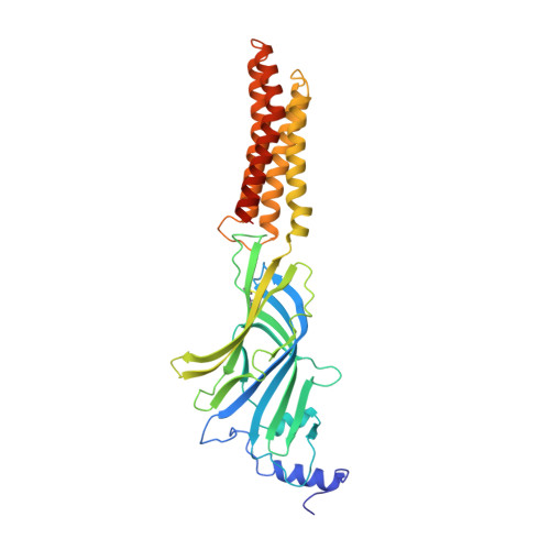
Crystal Structure of a Human Gabaa Receptor

Miller, P.S.Aricescu, A.R.

(2014) Nature 512: 270

  • DOI: https://doi.org/10.1038/nature13293
  • Primary Citation Related Structures: 
    4COF

  • PubMed Abstract: 

    Type-A γ-aminobutyric acid receptors (GABAARs) are the principal mediators of rapid inhibitory synaptic transmission in the human brain. A decline in GABAAR signalling triggers hyperactive neurological disorders such as insomnia, anxiety and epilepsy. Here we present the first three-dimensional structure of a GABAAR, the human β3 homopentamer, at 3 Å resolution. This structure reveals architectural elements unique to eukaryotic Cys-loop receptors, explains the mechanistic consequences of multiple human disease mutations and shows an unexpected structural role for a conserved N-linked glycan. The receptor was crystallized bound to a previously unknown agonist, benzamidine, opening a new avenue for the rational design of GABAAR modulators. The channel region forms a closed gate at the base of the pore, representative of a desensitized state. These results offer new insights into the signalling mechanisms of pentameric ligand-gated ion channels and enhance current understanding of GABAergic neurotransmission.


  • Organizational Affiliation
    • Division of Structural Biology, Wellcome Trust Centre for Human Genetics, University of Oxford, Roosevelt Drive, Oxford OX3 7BN, UK.

Macromolecule Content 

  • Total Structure Weight: 209.2 kDa 
  • Atom Count: 13,972 
  • Modeled Residue Count: 1,665 
  • Deposited Residue Count: 1,775 
  • Unique protein chains: 1

Macromolecules

Find similar proteins by:|  3D Structure
Entity ID: 1
MoleculeChains  Sequence LengthOrganismDetailsImage
GAMMA-AMINOBUTYRIC ACID RECEPTOR SUBUNIT BETA-3
A, B, C, D, E
355Homo sapiensMutation(s): 0 
Membrane Entity: Yes 
UniProt & NIH Common Fund Data Resources
Find proteins for P28472 (Homo sapiens)
Explore P28472 
Go to UniProtKB:  P28472
PHAROS:  P28472
GTEx:  ENSG00000166206 
Entity Groups
Sequence Clusters30% Identity50% Identity70% Identity90% Identity95% Identity100% Identity
UniProt GroupP28472
Glycosylation
Glycosylation Sites: 3Go to GlyGen: P28472-1
Sequence Annotations
Expand
Reference Sequence

Oligosaccharides

Help  
Entity ID: 2
MoleculeChains Length2D Diagram GlycosylationD Interactions
beta-D-mannopyranose-(1-4)-2-acetamido-2-deoxy-beta-D-glucopyranose-(1-4)-2-acetamido-2-deoxy-beta-D-glucopyranose
F, G, H, I, J
3N-Glycosylation
Glycosylation Resources
GlyTouCan: G15407YE
GlyCosmos: G15407YE
GlyGen: G15407YE

Small Molecules

Ligands 3 Unique
IDChains Name / Formula / InChI Key2D Diagram3D Interactions
NAG

Query on NAG


Download:Ideal Coordinates CCD File
L [auth A]
N [auth A]
Q [auth B]
T [auth C]
W [auth D]
L [auth A],
N [auth A],
Q [auth B],
T [auth C],
W [auth D],
Z [auth E]
2-acetamido-2-deoxy-beta-D-glucopyranose
C8 H15 N O6
OVRNDRQMDRJTHS-FMDGEEDCSA-N
BEN

Query on BEN


Download:Ideal Coordinates CCD File
K [auth A],
O [auth B],
R [auth C],
U [auth D],
X [auth E]
BENZAMIDINE
C7 H8 N2
PXXJHWLDUBFPOL-UHFFFAOYSA-N
CL

Query on CL


Download:Ideal Coordinates CCD File
M [auth A],
P [auth B],
S [auth C],
V [auth D],
Y [auth E]
CHLORIDE ION
Cl
VEXZGXHMUGYJMC-UHFFFAOYSA-M

Experimental Data & Validation

Experimental Data

  • Method: X-RAY DIFFRACTION
  • Resolution: 2.97 Å
  • R-Value Free:  0.226 (Depositor), 0.235 (DCC) 
  • R-Value Work:  0.205 (Depositor), 0.215 (DCC) 
  • R-Value Observed: 0.206 (Depositor) 
Space Group: C 1 2 1
Unit Cell:
Length ( Å )Angle ( ˚ )
a = 174.1α = 90
b = 108.9β = 107.43
c = 207.44γ = 90
Software Package:
Software NamePurpose
BUSTERrefinement
XDSdata reduction
SCALAdata scaling
PHASERphasing

Structure Validation

View Full Validation Report


Ligand Structure Quality Assessment 


Entry History 

Deposition Data

Revision History  (Full details and data files)

  • Version 1.0: 2014-06-04
    Type: Initial release
  • Version 1.1: 2014-06-11
    Changes: Structure summary
  • Version 1.2: 2014-06-18
    Changes: Database references
  • Version 1.3: 2014-08-27
    Changes: Database references
  • Version 2.0: 2020-07-29
    Type: Remediation
    Reason: Carbohydrate remediation
    Changes: Atomic model, Data collection, Derived calculations, Other, Structure summary
  • Version 2.1: 2023-12-20
    Changes: Data collection, Database references, Derived calculations, Refinement description, Structure summary
  • Version 2.2: 2024-11-06
    Changes: Structure summary