4CMR | pdb_00004cmr

The crystal structure of novel exo-type maltose-forming amylase(Py04_0872) from Pyrococcus sp. ST04


Experimental Data Snapshot

  • Method: X-RAY DIFFRACTION
  • Resolution: 1.80 Å
  • R-Value Free: 
    0.225 (Depositor), 0.220 (DCC) 
  • R-Value Work: 
    0.198 (Depositor), 0.193 (DCC) 
  • R-Value Observed: 
    0.198 (Depositor) 

wwPDB Validation 3D Report Full Report

Validation slider image for 4CMR

This is version 1.3 of the entry. See complete history

Literature

Structural Features Underlying the Selective Cleavage of a Novel Exo-Type Maltose-Forming Amylase from Pyrococcus Sp. St04

Park, K.-H.Jung, J.-H.Park, S.-G.Lee, M.-E.Holden, J.F.Park, C.-S.Woo, E.-J.

(2014) Acta Crystallogr D Biol Crystallogr 70: 1659

  • DOI: https://doi.org/10.1107/S1399004714006567
  • Primary Citation Related Structures: 
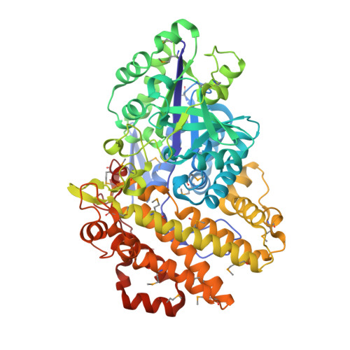
    4CMR

  • PubMed Abstract: 

    A novel maltose-forming α-amylase (PSMA) was recently found in the hyperthermophilic archaeon Pyrococcus sp. ST04. This enzyme shows <13% amino-acid sequence identity to other known α-amylases and displays a unique enzymatic property in that it hydrolyzes both α-1,4-glucosidic and α-1,6-glucosidic linkages of substrates, recognizing only maltose units, in an exo-type manner. Here, the crystal structure of PSMA at a resolution of 1.8 Å is reported, showing a tight ring-shaped tetramer with monomers composed of two domains: an N-domain (amino acids 1-341) with a typical GH57 family (β/α)7-barrel fold and a C-domain (amino acids 342-597) composed of α-helical bundles. A small closed cavity observed in proximity to the catalytic residues Glu153 and Asp253 at the domain interface has the appropriate volume and geometry to bind a maltose unit, accounting for the selective exo-type maltose hydrolysis of the enzyme. A narrow gate at the putative subsite +1 formed by residue Phe218 and Phe452 is essential for specific cleavage of glucosidic bonds. The closed cavity at the active site is connected to a short substrate-binding channel that extends to the central hole of the tetramer, exhibiting a geometry that is significantly different from classical maltogenic amylases or β-amylases. The structural features of this novel exo-type maltose-forming α-amylase provide a molecular basis for its unique enzymatic characteristics and for its potential use in industrial applications and protein engineering.


  • Organizational Affiliation
    • Korea Research Institute of Bioscience and Biotechnology (KRIBB), Daejeon 305-806, Republic of Korea.

Macromolecule Content 

  • Total Structure Weight: 142.16 kDa 
  • Atom Count: 10,845 
  • Modeled Residue Count: 1,192 
  • Deposited Residue Count: 1,194 
  • Unique protein chains: 1

Macromolecules

Find similar proteins by:|  3D Structure
Entity ID: 1
MoleculeChains  Sequence LengthOrganismDetailsImage
GLYCOSYL HYDROLASE/DEACETYLASE FAMILY PROTEIN
A, B
597Pyrococcus sp. ST04Mutation(s): 0 
UniProt
Find proteins for A0ACD6B8M0 (Pyrococcus sp. ST04)
Explore A0ACD6B8M0 
Go to UniProtKB:  A0ACD6B8M0
Entity Groups
Sequence Clusters30% Identity50% Identity70% Identity90% Identity95% Identity100% Identity
UniProt GroupA0ACD6B8M0
Sequence Annotations
Expand
Reference Sequence

Small Molecules

Modified Residues  1 Unique
IDChains TypeFormula2D DiagramParent
MSE
Query on MSE
A, B
L-PEPTIDE LINKINGC5 H11 N O2 SeMET

Experimental Data & Validation

Experimental Data

  • Method: X-RAY DIFFRACTION
  • Resolution: 1.80 Å
  • R-Value Free:  0.225 (Depositor), 0.220 (DCC) 
  • R-Value Work:  0.198 (Depositor), 0.193 (DCC) 
  • R-Value Observed: 0.198 (Depositor) 
Space Group: C 2 2 21
Unit Cell:
Length ( Å )Angle ( ˚ )
a = 117.639α = 90
b = 370.794β = 90
c = 82.135γ = 90
Software Package:
Software NamePurpose
PHENIXrefinement
HKL-2000data reduction
HKL-2000data scaling
PHENIXphasing

Structure Validation

View Full Validation Report


Entry History 

Deposition Data

Revision History  (Full details and data files)

  • Version 1.0: 2014-10-22
    Type: Initial release
  • Version 1.1: 2015-09-23
    Changes: Data collection
  • Version 1.2: 2019-10-16
    Changes: Data collection, Derived calculations, Experimental preparation, Other
  • Version 1.3: 2024-10-16
    Changes: Data collection, Database references, Structure summary