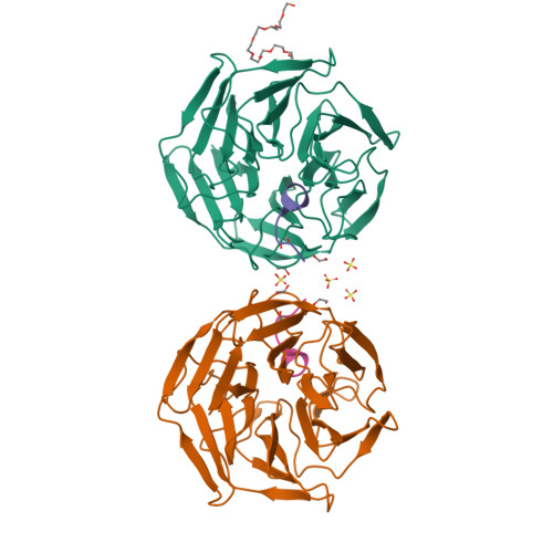
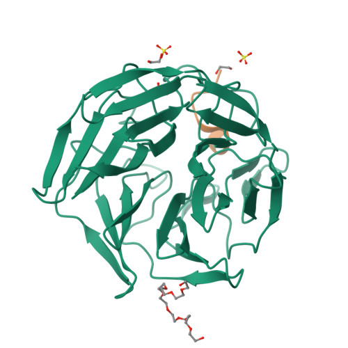
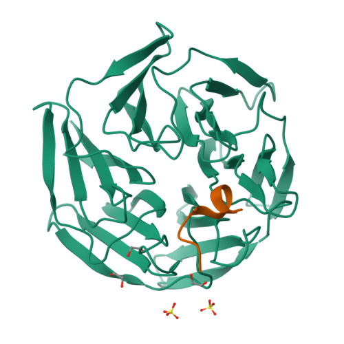
4CHB

Crystal structure of the human KLHL2 Kelch domain in complex with a WNK4 peptide


Experimental Data Snapshot

  • Method: X-RAY DIFFRACTION
  • Resolution: 1.56 Å
  • R-Value Free: 0.194 
  • R-Value Work: 0.177 
  • R-Value Observed: 0.178 

Starting Model: experimental
View more details

wwPDB Validation   3D Report Full Report


This is version 1.7 of the entry. See complete history


Literature

Structural and Biochemical Characterisation of the Klhl3-Wnk Kinase Interaction Important in Blood Pressure Regulation.

Schumacher, F.Sorrell, F.J.Alessi, D.R.Bullock, A.N.Kurz, T.

(2014) Biochem J 460: 237

  • DOI: https://doi.org/10.1042/BJ20140153
  • Primary Citation of Related Structures:  
    4CH9, 4CHB

  • PubMed Abstract: 

    WNK1 [with no lysine (K)] and WNK4 regulate blood pressure by controlling the activity of ion co-transporters in the kidney. Groundbreaking work has revealed that the ubiquitylation and hence levels of WNK isoforms are controlled by a Cullin-RING E3 ubiquitin ligase complex (CRL3KLHL3) that utilizes CUL3 (Cullin3) and its substrate adaptor, KLHL3 (Kelch-like protein 3). Loss-of-function mutations in either CUL3 or KLHL3 cause the hereditary high blood pressure disease Gordon's syndrome by stabilizing WNK isoforms. KLHL3 binds to a highly conserved degron motif located within the C-terminal non-catalytic domain of WNK isoforms. This interaction is essential for ubiquitylation by CRL3KLHL3 and disease-causing mutations in WNK4 and KLHL3 exert their effects on blood pressure by disrupting this interaction. In the present study, we report on the crystal structure of the KLHL3 Kelch domain in complex with the WNK4 degron motif. This reveals an intricate web of interactions between conserved residues on the surface of the Kelch domain β-propeller and the WNK4 degron motif. Importantly, many of the disease-causing mutations inhibit binding by disrupting critical interface contacts. We also present the structure of the WNK4 degron motif in complex with KLHL2 that has also been reported to bind WNK4. This confirms that KLHL2 interacts with WNK kinases in a similar manner to KLHL3, but strikingly different to how another KLHL protein, KEAP1 (Kelch-like enoyl-CoA hydratase-associated protein 1), binds to its substrate NRF2 (nuclear factor-erythroid 2-related factor 2). The present study provides further insights into how Kelch-like adaptor proteins recognize their substrates and provides a structural basis for how mutations in WNK4 and KLHL3 lead to hypertension.


  • Organizational Affiliation

    *MRC Protein Phosphorylation and Ubiquitylation Unit, College of Life Sciences, University of Dundee, Dow Street, Dundee DD1 5EH, Scotland, U.K.


Macromolecules
Find similar proteins by:  (by identity cutoff)  |  3D Structure
Entity ID: 1
MoleculeChains Sequence LengthOrganismDetailsImage
KELCH-LIKE PROTEIN 2
A, B
302Homo sapiensMutation(s): 0 
UniProt & NIH Common Fund Data Resources
Find proteins for O95198 (Homo sapiens)
Explore O95198 
Go to UniProtKB:  O95198
PHAROS:  O95198
GTEx:  ENSG00000109466 
Entity Groups  
Sequence Clusters30% Identity50% Identity70% Identity90% Identity95% Identity100% Identity
UniProt GroupO95198
Sequence Annotations
Expand
  • Reference Sequence

Find similar proteins by:  Sequence   |   3D Structure  

Entity ID: 2
MoleculeChains Sequence LengthOrganismDetailsImage
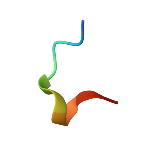
SERINE/THREONINE-PROTEIN KINASE WNK4
C, D
11Homo sapiensMutation(s): 0 
EC: 2.7.11.1
UniProt & NIH Common Fund Data Resources
Find proteins for Q96J92 (Homo sapiens)
Explore Q96J92 
Go to UniProtKB:  Q96J92
PHAROS:  Q96J92
GTEx:  ENSG00000126562 
Entity Groups  
Sequence Clusters30% Identity50% Identity70% Identity90% Identity95% Identity100% Identity
UniProt GroupQ96J92
Sequence Annotations
Expand
  • Reference Sequence
Small Molecules
Ligands 3 Unique
IDChains Name / Formula / InChI Key2D Diagram3D Interactions
12P
Query on 12P

Download Ideal Coordinates CCD File 
H [auth A]DODECAETHYLENE GLYCOL
C24 H50 O13
WRZXKWFJEFFURH-UHFFFAOYSA-N
SO4
Query on SO4

Download Ideal Coordinates CCD File 
F [auth A],
I [auth A],
J [auth B],
M [auth B]
SULFATE ION
O4 S
QAOWNCQODCNURD-UHFFFAOYSA-L
EDO
Query on EDO

Download Ideal Coordinates CCD File 
E [auth A]
G [auth A]
K [auth B]
L [auth B]
N [auth C]
E [auth A],
G [auth A],
K [auth B],
L [auth B],
N [auth C],
O [auth D]
1,2-ETHANEDIOL
C2 H6 O2
LYCAIKOWRPUZTN-UHFFFAOYSA-N
Experimental Data & Validation

Experimental Data

  • Method: X-RAY DIFFRACTION
  • Resolution: 1.56 Å
  • R-Value Free: 0.194 
  • R-Value Work: 0.177 
  • R-Value Observed: 0.178 
  • Space Group: P 43 21 2
Unit Cell:
Length ( Å )Angle ( ˚ )
a = 117.7α = 90
b = 117.7β = 90
c = 106.65γ = 90
Software Package:
Software NamePurpose
PHENIXrefinement
MOSFLMdata reduction
SCALAdata scaling
PHASERphasing

Structure Validation

View Full Validation Report


Entry History 

Revision History  (Full details and data files)

  • Version 1.0: 2014-01-08
    Type: Initial release
  • Version 1.1: 2014-05-21
    Changes: Database references
  • Version 1.2: 2014-05-28
    Changes: Database references
  • Version 1.3: 2015-07-22
    Changes: Structure summary
  • Version 1.4: 2018-01-24
    Changes: Structure summary
  • Version 1.5: 2019-03-06
    Changes: Data collection, Experimental preparation
  • Version 1.6: 2019-05-29
    Changes: Data collection, Experimental preparation
  • Version 1.7: 2023-12-20
    Changes: Data collection, Database references, Derived calculations, Other, Refinement description