4CDP

Improved coordinates for Escherichia coli O157:H7 heme degrading enzyme ChuS.


Experimental Data Snapshot

  • Method: X-RAY DIFFRACTION
  • Resolution: 1.45 Å
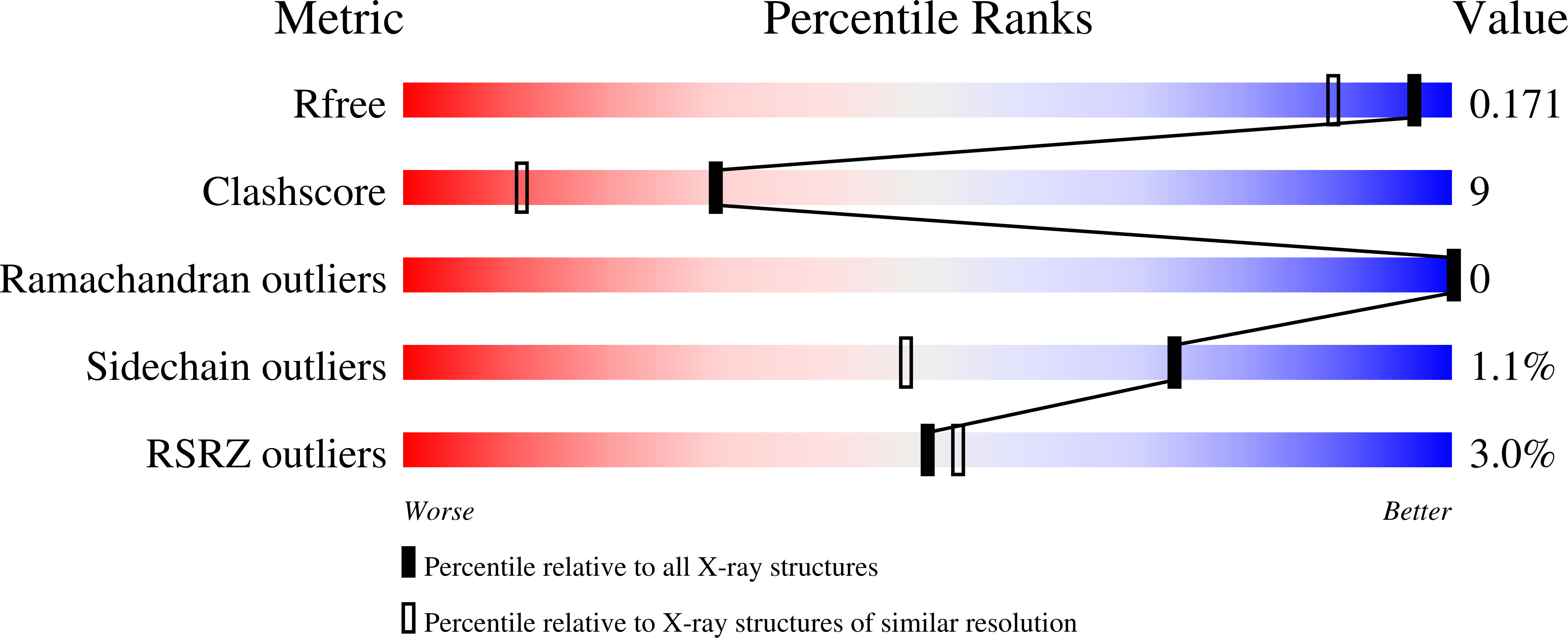
  • R-Value Free: 0.173 
  • R-Value Work: 0.133 
  • R-Value Observed: 0.135 

Starting Model: experimental
View more details

wwPDB Validation   3D Report Full Report


Ligand Structure Quality Assessment 


This is version 1.3 of the entry. See complete history


Literature

Structure of the Escherichia Coli O157:H7 Heme Oxygenase Chus in Complex with Heme and Enzymatic Inactivation by Mutation of the Heme Coordinating Residue His-193.

Suits, M.D.Jaffer, N.Jia, Z.

(2006) J Biol Chem 281: 36776

  • DOI: https://doi.org/10.1074/jbc.M607684200
  • Primary Citation of Related Structures:  
    4CDP

  • PubMed Abstract: 

    Heme oxygenases catalyze the oxidation of heme to biliverdin, CO, and free iron. For pathogenic microorganisms, heme uptake and degradation are critical mechanisms for iron acquisition that enable multiplication and survival within hosts they invade. Here we report the first crystal structure of the pathogenic Escherichia coli O157:H7 heme oxygenase ChuS in complex with heme at 1.45 A resolution. When compared with other heme oxygenases, ChuS has a unique fold, including structural repeats and a beta-sheet core. Not surprisingly, the mode of heme coordination by ChuS is also distinct, whereby heme is largely stabilized by residues from the C-terminal domain, assisted by a distant arginine from the N-terminal domain. Upon heme binding, there is no large conformational change beyond the fine tuning of a key histidine (His-193) residue. Most intriguingly, in contrast to other heme oxygenases, the propionic side chains of heme are orientated toward the protein core, exposing the alpha-meso carbon position where O(2) is added during heme degradation. This unique orientation may facilitate presentation to an electron donor, explaining the significantly reduced concentration of ascorbic acid needed for the reaction. Based on the ChuS-heme structure, we converted the histidine residue responsible for axial coordination of the heme group to an asparagine residue (H193N), as well as converting a second histidine to an alanine residue (H73A) for comparison purposes. We employed spectral analysis and CO measurement by gas chromatography to analyze catalysis by ChuS, H193N, and H73A, demonstrating that His-193 is the key residue for the heme-degrading activity of ChuS.


  • Organizational Affiliation

    Department of Biochemistry, Queen's University, Kingston, Ontario K7L 3N6, Canada.


Macromolecules
Find similar proteins by:  (by identity cutoff)  |  3D Structure
Entity ID: 1
MoleculeChains Sequence LengthOrganismDetailsImage
PUTATIVE HEME/HEMOGLOBIN TRANSPORT PROTEIN354Escherichia coli O157:H7Mutation(s): 0 
UniProt
Find proteins for Q8X5N8 (Escherichia coli O157:H7)
Explore Q8X5N8 
Go to UniProtKB:  Q8X5N8
Entity Groups  
Sequence Clusters30% Identity50% Identity70% Identity90% Identity95% Identity100% Identity
UniProt GroupQ8X5N8
Sequence Annotations
Expand
  • Reference Sequence
Small Molecules
Ligands 3 Unique
IDChains Name / Formula / InChI Key2D Diagram3D Interactions
HEM
Query on HEM

Download Ideal Coordinates CCD File 
C [auth A]PROTOPORPHYRIN IX CONTAINING FE
C34 H32 Fe N4 O4
KABFMIBPWCXCRK-RGGAHWMASA-L
EDO
Query on EDO

Download Ideal Coordinates CCD File 
D [auth A]
E [auth A]
F [auth A]
G [auth A]
H [auth A]
D [auth A],
E [auth A],
F [auth A],
G [auth A],
H [auth A],
I [auth A],
J [auth A],
K [auth A],
L [auth A],
M [auth A],
N [auth A],
O [auth A]
1,2-ETHANEDIOL
C2 H6 O2
LYCAIKOWRPUZTN-UHFFFAOYSA-N
FMT
Query on FMT

Download Ideal Coordinates CCD File 
B [auth A]FORMIC ACID
C H2 O2
BDAGIHXWWSANSR-UHFFFAOYSA-N
Experimental Data & Validation

Experimental Data

  • Method: X-RAY DIFFRACTION
  • Resolution: 1.45 Å
  • R-Value Free: 0.173 
  • R-Value Work: 0.133 
  • R-Value Observed: 0.135 
  • Space Group: H 3
Unit Cell:
Length ( Å )Angle ( ˚ )
a = 106.487α = 90
b = 106.487β = 90
c = 90.229γ = 120
Software Package:
Software NamePurpose
REFMACrefinement
HKL-2000data reduction
SCALEPACKdata scaling
PHASERphasing

Structure Validation

View Full Validation Report


Ligand Structure Quality Assessment 


Entry History 

Deposition Data

Revision History  (Full details and data files)

  • Version 1.0: 2013-11-13
    Type: Initial release
  • Version 1.1: 2019-01-30
    Changes: Data collection, Experimental preparation
  • Version 1.2: 2019-03-06
    Changes: Data collection, Experimental preparation
  • Version 1.3: 2023-12-20
    Changes: Data collection, Database references, Derived calculations, Other, Refinement description