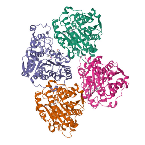
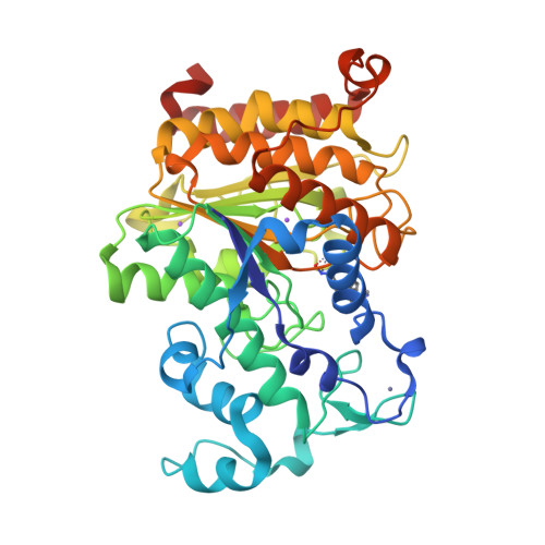
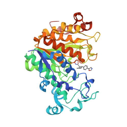
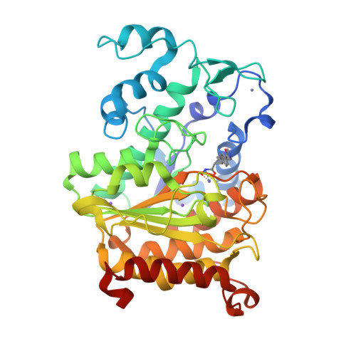
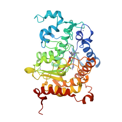
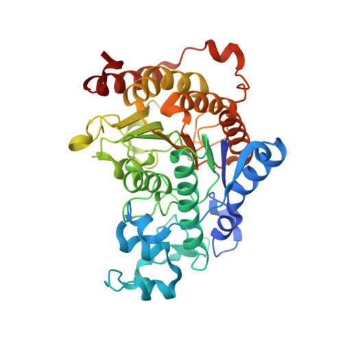
4CBY

Design, synthesis, and biological evaluation of potent and selective Class IIa HDAC inhibitors as a potential therapy for Huntington's disease


Experimental Data Snapshot

  • Method: X-RAY DIFFRACTION
  • Resolution: 2.72 Å
  • R-Value Free: 0.246 
  • R-Value Work: 0.209 
  • R-Value Observed: 0.211 

wwPDB Validation   3D Report Full Report


Ligand Structure Quality Assessment 


This is version 1.3 of the entry. See complete history


Literature

Design, synthesis, and biological evaluation of potent and selective class IIa histone deacetylase (HDAC) inhibitors as a potential therapy for Huntington's disease.

Burli, R.W.Luckhurst, C.A.Aziz, O.Matthews, K.L.Yates, D.Lyons, K.A.Beconi, M.McAllister, G.Breccia, P.Stott, A.J.Penrose, S.D.Wall, M.Lamers, M.Leonard, P.Muller, I.Richardson, C.M.Jarvis, R.Stones, L.Hughes, S.Wishart, G.Haughan, A.F.O'Connell, C.Mead, T.McNeil, H.Vann, J.Mangette, J.Maillard, M.Beaumont, V.Munoz-Sanjuan, I.Dominguez, C.

(2013) J Med Chem 56: 9934-9954

  • DOI: https://doi.org/10.1021/jm4011884
  • Primary Citation of Related Structures:  
    4CBT, 4CBY

  • PubMed Abstract: 

    Inhibition of class IIa histone deacetylase (HDAC) enzymes have been suggested as a therapeutic strategy for a number of diseases, including Huntington's disease. Catalytic-site small molecule inhibitors of the class IIa HDAC4, -5, -7, and -9 were developed. These trisubstituted diarylcyclopropanehydroxamic acids were designed to exploit a lower pocket that is characteristic for the class IIa HDACs, not present in other HDAC classes. Selected inhibitors were cocrystallized with the catalytic domain of human HDAC4. We describe the first HDAC4 catalytic domain crystal structure in a "closed-loop" form, which in our view represents the biologically relevant conformation. We have demonstrated that these molecules can differentiate class IIa HDACs from class I and class IIb subtypes. They exhibited pharmacokinetic properties that should enable the assessment of their therapeutic benefit in both peripheral and CNS disorders. These selective inhibitors provide a means for evaluating potential efficacy in preclinical models in vivo.


  • Organizational Affiliation

    BioFocus , Chesterford Research Park, Saffron Walden, Essex, CB10 1XL, United Kingdom.


Macromolecules
Find similar proteins by:  (by identity cutoff)  |  3D Structure
Entity ID: 1
MoleculeChains Sequence LengthOrganismDetailsImage
HISTONE DEACETYLASE 4
A, B, C, D
395Homo sapiensMutation(s): 1 
EC: 3.5.1.98
UniProt & NIH Common Fund Data Resources
Find proteins for P56524 (Homo sapiens)
Go to UniProtKB:  P56524
PHAROS:  P56524
GTEx:  ENSG00000068024 
Sequence Annotations
Expand
  • Reference Sequence
Small Molecules
Ligands 3 Unique
IDChains Name / Formula / InChI Key2D Diagram3D Interactions
KEE
Query on KEE

Download Ideal Coordinates CCD File 
E [auth A],
J [auth B],
O [auth C],
T [auth D]
(1R,2R,3R)-2-[4-(1,3-oxazol-5-yl)phenyl]-N-oxidanyl-3-phenyl-cyclopropane-1-carboxamide
C19 H16 N2 O3
PYWCXCBMYMFPTR-KZNAEPCWSA-N
ZN
Query on ZN

Download Ideal Coordinates CCD File 
F [auth A]
G [auth A]
K [auth B]
L [auth B]
P [auth C]
F [auth A],
G [auth A],
K [auth B],
L [auth B],
P [auth C],
Q [auth C],
U [auth D],
V [auth D]
ZINC ION
Zn
PTFCDOFLOPIGGS-UHFFFAOYSA-N
NA
Query on NA

Download Ideal Coordinates CCD File 
H [auth A]
I [auth A]
M [auth B]
N [auth B]
R [auth C]
H [auth A],
I [auth A],
M [auth B],
N [auth B],
R [auth C],
S [auth C],
W [auth D],
X [auth D]
SODIUM ION
Na
FKNQFGJONOIPTF-UHFFFAOYSA-N
Binding Affinity Annotations 
IDSourceBinding Affinity
KEE PDBBind:  4CBY IC50: 20 (nM) from 1 assay(s)
BindingDB:  4CBY IC50: min: 20, max: 200 (nM) from 2 assay(s)
Experimental Data & Validation

Experimental Data

  • Method: X-RAY DIFFRACTION
  • Resolution: 2.72 Å
  • R-Value Free: 0.246 
  • R-Value Work: 0.209 
  • R-Value Observed: 0.211 
  • Space Group: P 1 21 1
Unit Cell:
Length ( Å )Angle ( ˚ )
a = 54.151α = 90
b = 161.816β = 97.03
c = 83.732γ = 90
Software Package:
Software NamePurpose
REFMACrefinement
PHASERphasing

Structure Validation

View Full Validation Report


Ligand Structure Quality Assessment 


Entry History 

Revision History  (Full details and data files)

  • Version 1.0: 2013-12-11
    Type: Initial release
  • Version 1.1: 2014-01-15
    Changes: Database references
  • Version 1.2: 2018-02-07
    Changes: Database references, Structure summary
  • Version 1.3: 2024-05-08
    Changes: Data collection, Database references, Derived calculations, Other