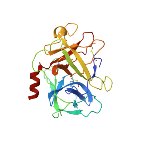
Ensemble Refinement Shows Conformational Flexibility in Crystal Structures of Human Complement Factor D
Forneris, F., Burnley, B.T., Gros, P.(2014) Acta Crystallogr D Biol Crystallogr 70: 733
- PubMed: 24598742
- DOI: https://doi.org/10.1107/S1399004713032549
- Primary Citation of Related Structures:
4CBN, 4CBO - PubMed Abstract:
Human factor D (FD) is a self-inhibited thrombin-like serine proteinase that is critical for amplification of the complement immune response. FD is activated by its substrate through interactions outside the active site. The substrate-binding, or `exosite', region displays a well defined and rigid conformation in FD. In contrast, remarkable flexibility is observed in thrombin and related proteinases, in which Na(+) and ligand binding is implied in allosteric regulation of enzymatic activity through protein dynamics. Here, ensemble refinement (ER) of FD and thrombin crystal structures is used to evaluate structure and dynamics simultaneously. A comparison with previously published NMR data for thrombin supports the ER analysis. The R202A FD variant has enhanced activity towards artificial peptides and simultaneously displays active and inactive conformations of the active site. ER revealed pronounced disorder in the exosite loops for this FD variant, reminiscent of thrombin in the absence of the stabilizing Na(+) ion. These data indicate that FD exhibits conformational dynamics like thrombin, but unlike in thrombin a mechanism has evolved in FD that locks the unbound native state into an ordered inactive conformation via the self-inhibitory loop. Thus, ensemble refinement of X-ray crystal structures may represent an approach alternative to spectroscopy to explore protein dynamics in atomic detail.
Organizational Affiliation:
Crystal and Structural Chemistry, Bijvoet Center for Biomolecular Research, Department of Chemistry, Faculty of Science, Utrecht University, Padualaan 8, 3584 CH Utrecht, The Netherlands.


