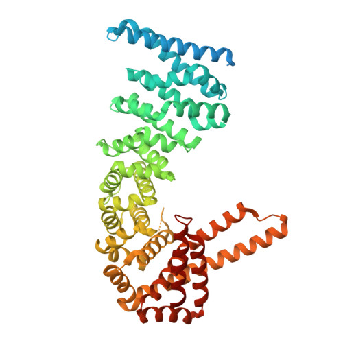
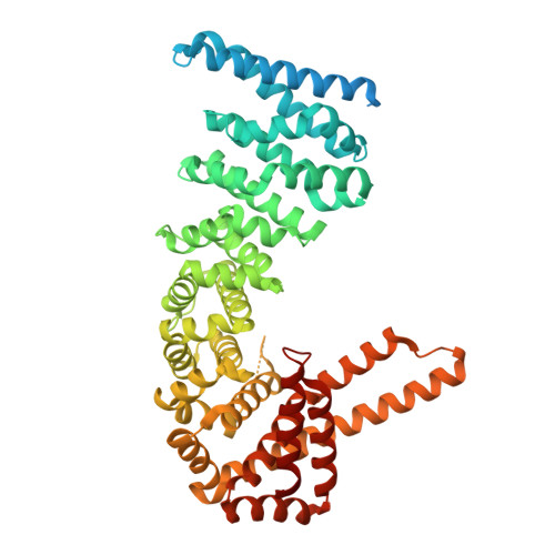
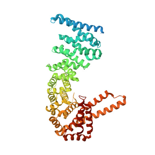
4CB9

Structure of full-length CTNNBL1 in P43212 space group


Experimental Data Snapshot

  • Method: X-RAY DIFFRACTION
  • Resolution: 3.00 Å
  • R-Value Free: 0.299 
  • R-Value Work: 0.218 
  • R-Value Observed: 0.222 

wwPDB Validation   3D Report Full Report


This is version 1.2 of the entry. See complete history


Literature

Structural and Mutational Analysis Reveals that Ctnnbl1 Binds Nlss in a Manner Distinct from that of its Closest Armadillo-Relative, Karyopherin Alpha

Ganesh, K.Maldegem, F.V.Telerman, S.B.Simpson, P.Johnson, C.M.Williams, R.L.Neuberger, M.S.Rada, C.

(2014) FEBS Lett 588: 21

  • DOI: https://doi.org/10.1016/j.febslet.2013.11.013
  • Primary Citation of Related Structures:  
    4CB8, 4CB9, 4CBA

  • PubMed Abstract: 

    CTNNBL1 is a spliceosome-associated protein that binds nuclear localization signals (NLSs) in splice factors CDC5L and Prp31 as well as the antibody diversifying enzyme AID. Here, crystal structures of human CTNNBL1 reveal a distinct structure from its closest homologue karyopherin-α. CTNNBL1 comprises a HEAT-like domain (including a nuclear export signal), a central armadillo domain, and a coiled-coil C-terminal domain. Structure-guided mutations of the region homologous to the karyopherin-α NLS-binding site fail to disrupt CTNNBL1-NLS interactions. Our results identify CTNNBL1 as a unique selective NLS-binding protein with striking differences from karyopherin-αs.


  • Organizational Affiliation

    Medical Research Council Laboratory of Molecular Biology, Francis Crick Avenue, Cambridge CB2 0QH, UK.


Macromolecules
Find similar proteins by:  (by identity cutoff)  |  3D Structure
Entity ID: 1
MoleculeChains Sequence LengthOrganismDetailsImage
BETA-CATENIN-LIKE PROTEIN 1571Homo sapiensMutation(s): 0 
UniProt & NIH Common Fund Data Resources
Find proteins for Q8WYA6 (Homo sapiens)
Explore Q8WYA6 
Go to UniProtKB:  Q8WYA6
PHAROS:  Q8WYA6
GTEx:  ENSG00000132792 
Entity Groups  
Sequence Clusters30% Identity50% Identity70% Identity90% Identity95% Identity100% Identity
UniProt GroupQ8WYA6
Sequence Annotations
Expand
  • Reference Sequence
Experimental Data & Validation

Experimental Data

  • Method: X-RAY DIFFRACTION
  • Resolution: 3.00 Å
  • R-Value Free: 0.299 
  • R-Value Work: 0.218 
  • R-Value Observed: 0.222 
  • Space Group: P 43 21 2
Unit Cell:
Length ( Å )Angle ( ˚ )
a = 66.727α = 90
b = 66.727β = 90
c = 325.883γ = 90
Software Package:
Software NamePurpose
XDSdata reduction
Aimlessdata scaling
PHASERphasing
SHARPphasing
REFMACrefinement

Structure Validation

View Full Validation Report


Entry History 

Deposition Data

Revision History  (Full details and data files)

  • Version 1.0: 2013-12-04
    Type: Initial release
  • Version 1.1: 2014-01-15
    Changes: Database references
  • Version 1.2: 2024-05-08
    Changes: Data collection, Database references, Other