4C9R

Xenopus ZNRF3 ectodomain in complex with Xenopus RSPO2 Fu1-Fu2 crystal form I


Experimental Data Snapshot

  • Method: X-RAY DIFFRACTION
  • Resolution: 2.10 Å
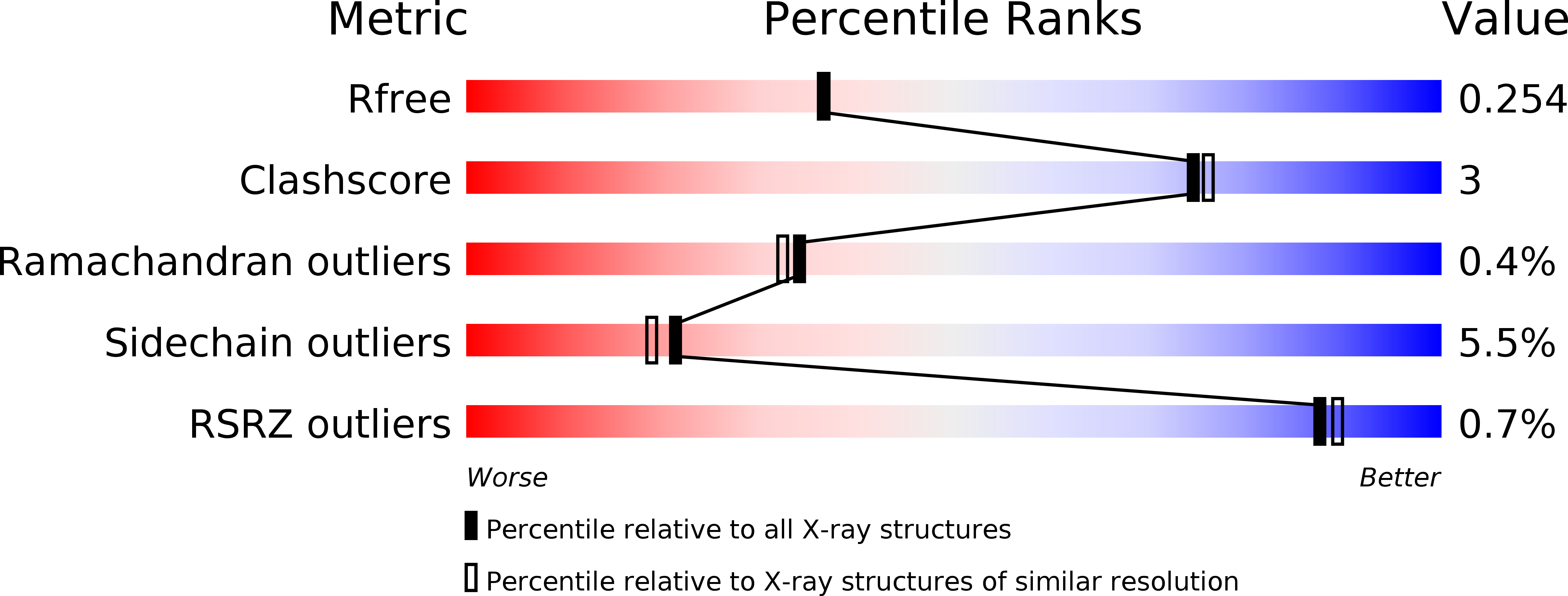
  • R-Value Free: 0.246 
  • R-Value Work: 0.188 
  • R-Value Observed: 0.190 

wwPDB Validation   3D Report Full Report


This is version 1.2 of the entry. See complete history


Literature

Structural and Molecular Basis of Znrf3/Rnf43 Transmembrane Ubiquitin Ligase Inhibition by the Wnt Agonist R-Spondin.

Zebisch, M.Xu, Y.Krastev, C.Macdonald, B.T.Chen, M.Gilbert, R.J.C.He, X.Jones, E.Y.

(2013) Nat Commun 4: 2787

  • DOI: https://doi.org/10.1038/ncomms3787
  • Primary Citation of Related Structures:  
    4C84, 4C85, 4C86, 4C8A, 4C8C, 4C8F, 4C8P, 4C8T, 4C8U, 4C8V, 4C8W, 4C99, 4C9A, 4C9E, 4C9R, 4C9U, 4C9V

  • PubMed Abstract: 

    The four R-spondin (Rspo) proteins are secreted agonists of Wnt signalling in vertebrates, functioning in embryogenesis and adult stem cell biology. Through ubiquitination and degradation of Wnt receptors, the transmembrane E3 ubiquitin ligase ZNRF3 and related RNF43 antagonize Wnt signalling. Rspo ligands have been reported to inhibit the ligase activity through direct interaction with ZNRF3 and RNF43. Here we report multiple crystal structures of the ZNRF3 ectodomain (ZNRF3(ecto)), a signalling-competent Furin1-Furin2 (Fu1-Fu2) fragment of Rspo2 (Rspo2(Fu1-Fu2)), and Rspo2(Fu1-Fu2) in complex with ZNRF3(ecto), or RNF43(ecto). A prominent loop in Fu1 clamps into equivalent grooves in the ZNRF3(ecto) and RNF43(ecto) surface. Rspo binding enhances dimerization of ZNRF3(ecto) but not of RNF43(ecto). Comparison of the four Rspo proteins, mutants and chimeras in biophysical and cellular assays shows that their signalling potency depends on their ability to recruit ZNRF3 or RNF43 via Fu1 into a complex with LGR receptors, which interact with Rspo via Fu2.


  • Organizational Affiliation

    Division of Structural Biology, Wellcome Trust Centre for Human Genetics, University of Oxford, Oxford OX3 7BN, UK.


Macromolecules
Find similar proteins by:  (by identity cutoff)  |  3D Structure
Entity ID: 1
MoleculeChains Sequence LengthOrganismDetailsImage
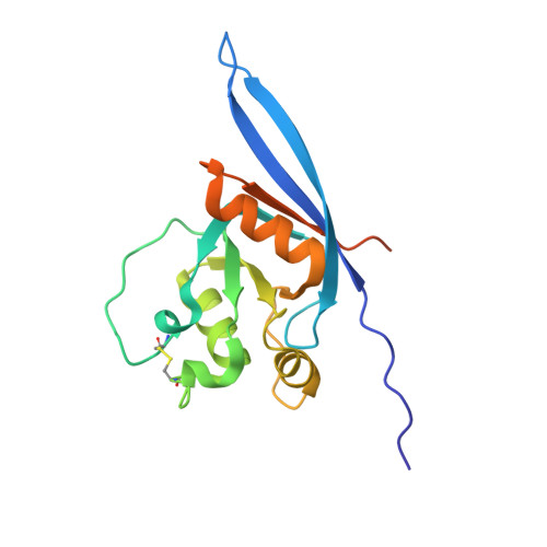
E3 UBIQUITIN-PROTEIN LIGASE ZNRF3
A, C
182Xenopus tropicalisMutation(s): 0 
EC: 6.3.2 (PDB Primary Data), 2.3.2.27 (UniProt)
UniProt
Find proteins for Q08D68 (Xenopus tropicalis)
Explore Q08D68 
Go to UniProtKB:  Q08D68
Entity Groups  
Sequence Clusters30% Identity50% Identity70% Identity90% Identity95% Identity100% Identity
UniProt GroupQ08D68
Sequence Annotations
Expand
  • Reference Sequence
Find similar proteins by:  (by identity cutoff)  |  3D Structure
Entity ID: 2
MoleculeChains Sequence LengthOrganismDetailsImage
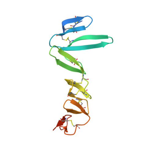
R-SPONDIN-2
B, D
121Xenopus tropicalisMutation(s): 0 
UniProt
Find proteins for Q5M7L6 (Xenopus tropicalis)
Explore Q5M7L6 
Go to UniProtKB:  Q5M7L6
Entity Groups  
Sequence Clusters30% Identity50% Identity70% Identity90% Identity95% Identity100% Identity
UniProt GroupQ5M7L6
Sequence Annotations
Expand
  • Reference Sequence
Experimental Data & Validation

Experimental Data

  • Method: X-RAY DIFFRACTION
  • Resolution: 2.10 Å
  • R-Value Free: 0.246 
  • R-Value Work: 0.188 
  • R-Value Observed: 0.190 
  • Space Group: P 1 21 1
Unit Cell:
Length ( Å )Angle ( ˚ )
a = 55.99α = 90
b = 81.229β = 113
c = 71.632γ = 90
Software Package:
Software NamePurpose
REFMACrefinement

Structure Validation

View Full Validation Report


Entry History 

Deposition Data

Revision History  (Full details and data files)

  • Version 1.0: 2013-11-20
    Type: Initial release
  • Version 1.1: 2013-11-27
    Changes: Database references
  • Version 1.2: 2024-11-06
    Changes: Data collection, Database references, Other, Structure summary