A Novel Membrane Sensor Controls the Localization and Arfgef Activity of Bacterial Ralf.
Folly-Klan, M., Alix, E., Stalder, D., Ray, P., Duarte, L.V., Delprato, A., Zeghouf, M., Antonny, B., Campanacci, V., Roy, C.R., Cherfils, J.(2013) PLoS Pathog 9: 3747
- PubMed: 24244168
- DOI: https://doi.org/10.1371/journal.ppat.1003747
- Primary Citation of Related Structures:
4C7P - PubMed Abstract:
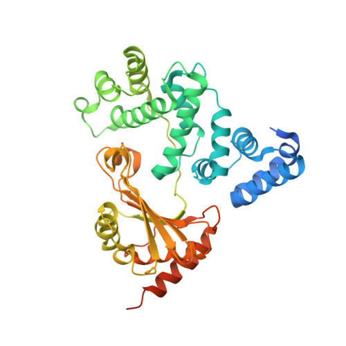
The intracellular bacterial pathogen Legionella pneumophila (Lp) evades destruction in macrophages by camouflaging in a specialized organelle, the Legionella-containing vacuole (LCV), where it replicates. The LCV maturates by incorporating ER vesicles, which are diverted by effectors that Lp injects to take control of host cell membrane transport processes. One of these effectors, RalF, recruits the trafficking small GTPase Arf1 to the LCV. LpRalF has a Sec7 domain related to host ArfGEFs, followed by a capping domain that intimately associates with the Sec7 domain to inhibit GEF activity. How RalF is activated to function as a LCV-specific ArfGEF is unknown. We combined the reconstitution of Arf activation on artificial membranes with cellular expression and Lp infection assays, to analyze how auto-inhibition is relieved for LpRalF to function in vivo. We find that membranes activate LpRalF by about 1000 fold, and identify the membrane-binding region as the region that inhibits the Sec7 active site. It is enriched in aromatic and positively charged residues, which establish a membrane sensor to control the GEF activity in accordance with specific lipid environments. A similar mechanism of activation is found in RalF from Rickettsia prowazekii (Rp), with a different aromatic/charged residues ratio that results in divergent membrane preferences. The membrane sensor is the primary determinant of the localization of LpRalF on the LCV, and drives the timing of Arf activation during infection. Finally, we identify a conserved motif in the capping domain, remote from the membrane sensor, which is critical for RalF activity presumably by organizing its active conformation. These data demonstrate that RalF proteins are regulated by a membrane sensor that functions as a binary switch to derepress ArfGEF activity when RalF encounters a favorable lipid environment, thus establishing a regulatory paradigm to ensure that Arf GTPases are efficiently activated at specific membrane locations.
Organizational Affiliation:
Laboratoire d'Enzymologie et Biochimie Structurales, Centre de Recherche de Gif, CNRS, Gif-sur-Yvette, France.


