4C55 | pdb_00004c55

Crystal structure of serum-derived human IgG4 Fc


Experimental Data Snapshot

  • Method: X-RAY DIFFRACTION
  • Resolution: 2.35 Å
  • R-Value Free: 
    0.238 (Depositor), 0.190 (DCC) 
  • R-Value Work: 
    0.188 (Depositor), 0.191 (DCC) 
  • R-Value Observed: 
    0.190 (Depositor) 

Starting Model: experimental
View more details

wwPDB Validation 3D Report Full Report

Validation slider image for 4C55

This is version 2.2 of the entry. See complete history

Literature

Structural Determinants of Unique Properties of Human Igg4-Fc

Davies, A.M.Rispens, T.Ooijevaar-Deheer, P.Gould, H.J.Jefferis, R.Aalberse, R.C.Sutton, B.J.

(2014) J Mol Biology 426: 630

  • DOI: https://doi.org/10.1016/j.jmb.2013.10.039
  • Primary Citation Related Structures: 
    4C54, 4C55

  • PubMed Abstract: 

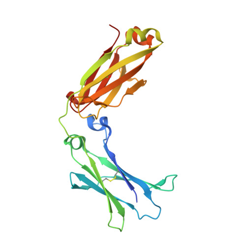
    Human IgG4, normally the least abundant of the four subclasses of IgG in serum, displays a number of unique biological properties. It can undergo heavy-chain exchange, also known as Fab-arm exchange, leading to the formation of monovalent but bispecific antibodies, and it interacts poorly with FcγRII and FcγRIII, and complement. These properties render IgG4 relatively "non-inflammatory" and have made it a suitable format for therapeutic monoclonal antibody production. However, IgG4 is also known to undergo Fc-mediated aggregation and has been implicated in auto-immune disease pathology. We report here the high-resolution crystal structures, at 1.9 and 2.35 Å, respectively, of human recombinant and serum-derived IgG4-Fc. These structures reveal conformational variability at the CH3-CH3 interface that may promote Fab-arm exchange, and a unique conformation for the FG loop in the CH2 domain that would explain the poor FcγRII, FcγRIII and C1q binding properties of IgG4 compared with IgG1 and -3. In contrast to other IgG subclasses, this unique conformation folds the FG loop away from the CH2 domain, precluding any interaction with the lower hinge region, which may further facilitate Fab-arm exchange by destabilisation of the hinge. The crystals of IgG4-Fc also display Fc-Fc packing contacts with very extensive interaction surfaces, involving both a consensus binding site in IgG-Fc at the CH2-CH3 interface and known hydrophobic aggregation motifs. These Fc-Fc interactions are compatible with intact IgG4 molecules and may provide a model for the formation of aggregates of IgG4 that can cause disease pathology in the absence of antigen.


  • Organizational Affiliation
    • Randall Division of Cell and Molecular Biophysics, King's College London, London SE1 1UL, United Kingdom; Medical Research Council and Asthma UK Centre in Allergic Mechanisms of Asthma, London SE1 9RT, United Kingdom. Electronic address: anna.davies@kcl.ac.uk.

Macromolecule Content 

  • Total Structure Weight: 52.97 kDa 
  • Atom Count: 3,499 
  • Modeled Residue Count: 414 
  • Deposited Residue Count: 436 
  • Unique protein chains: 1

Macromolecules

Find similar proteins by:|  3D Structure
Entity ID: 1
MoleculeChains  Sequence LengthOrganismDetailsImage
IG GAMMA-4 CHAIN C REGION
A, B
218Homo sapiensMutation(s): 0 
UniProt & NIH Common Fund Data Resources
Find proteins for P01861 (Homo sapiens)
Explore P01861 
Go to UniProtKB:  P01861
PHAROS:  P01861
GTEx:  ENSG00000211892 
Entity Groups
Sequence Clusters30% Identity50% Identity70% Identity90% Identity95% Identity100% Identity
UniProt GroupP01861
Glycosylation
Glycosylation Sites: 1Go to GlyGen: P01861-2
Sequence Annotations
Expand
Reference Sequence

Oligosaccharides

Help  
Entity ID: 2
MoleculeChains Length2D Diagram GlycosylationD Interactions
beta-D-galactopyranose-(1-4)-2-acetamido-2-deoxy-beta-D-glucopyranose-(1-2)-alpha-D-mannopyranose-(1-6)-[2-acetamido-2-deoxy-beta-D-glucopyranose-(1-2)-alpha-D-mannopyranose-(1-3)]beta-D-mannopyranose-(1-4)-2-acetamido-2-deoxy-beta-D-glucopyranose-(1-4)-[alpha-L-fucopyranose-(1-6)]2-acetamido-2-deoxy-beta-D-glucopyranose
C
9N-Glycosylation
Glycosylation Resources
GlyTouCan: G27919IH
GlyCosmos: G27919IH
GlyGen: G27919IH
Entity ID: 3
MoleculeChains Length2D Diagram GlycosylationD Interactions
beta-D-galactopyranose-(1-4)-2-acetamido-2-deoxy-beta-D-glucopyranose-(1-2)-alpha-D-mannopyranose-(1-6)-[2-acetamido-2-deoxy-beta-D-glucopyranose-(1-2)-alpha-D-mannopyranose-(1-3)]beta-D-mannopyranose-(1-4)-2-acetamido-2-deoxy-beta-D-glucopyranose-(1-4)-2-acetamido-2-deoxy-beta-D-glucopyranose
D
8N-Glycosylation
Glycosylation Resources
GlyTouCan: G89993FE
GlyCosmos: G89993FE
GlyGen: G89993FE

Experimental Data & Validation

Experimental Data

  • Method: X-RAY DIFFRACTION
  • Resolution: 2.35 Å
  • R-Value Free:  0.238 (Depositor), 0.190 (DCC) 
  • R-Value Work:  0.188 (Depositor), 0.191 (DCC) 
  • R-Value Observed: 0.190 (Depositor) 
Space Group: P 21 21 21
Unit Cell:
Length ( Å )Angle ( ˚ )
a = 74.298α = 90
b = 80.738β = 90
c = 99.19γ = 90
Software Package:
Software NamePurpose
PHENIXrefinement
XDSdata reduction
Aimlessdata scaling
MOLREPphasing

Structure Validation

View Full Validation Report


Entry History 

Deposition Data

Revision History  (Full details and data files)

  • Version 1.0: 2013-11-13
    Type: Initial release
  • Version 1.1: 2013-11-20
    Changes: Database references
  • Version 1.2: 2014-02-05
    Changes: Database references
  • Version 2.0: 2020-07-29
    Type: Remediation
    Reason: Carbohydrate remediation
    Changes: Advisory, Atomic model, Data collection, Derived calculations, Other, Structure summary
  • Version 2.1: 2023-12-20
    Changes: Data collection, Database references, Refinement description, Structure summary
  • Version 2.2: 2024-11-13
    Changes: Structure summary