4C51 | pdb_00004c51

Crystal Structure of the Catalase-Peroxidase (KatG) R418L mutant from Mycobacterium Tuberculosis


Experimental Data Snapshot

  • Method: X-RAY DIFFRACTION
  • Resolution: 3.10 Å
  • R-Value Free: 
    0.214 (Depositor), 0.214 (DCC) 
  • R-Value Work: 
    0.160 (Depositor), 0.161 (DCC) 
  • R-Value Observed: 
    0.163 (Depositor) 

Starting Model: experimental
View more details

wwPDB Validation 3D Report Full Report

Validation slider image for 4C51

Ligand Structure Quality Assessment 


This is version 1.4 of the entry. See complete history

Literature

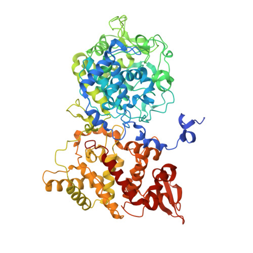
Access Channel Residues Ser315 and Asp137 in Mycobacterium Tuberculosis Catalase-Peroxidase (Katg) Control Peroxidatic Activation of the Pro-Drug Isoniazid.

Zhao, X.Hersleth, H.P.Zhu, J.Andersson, K.K.Magliozzo, R.S.

(2013) Chem Commun (Camb) 49: 11650

  • DOI: https://doi.org/10.1039/c3cc47022a
  • Primary Citation Related Structures: 
    4C50, 4C51

  • PubMed Abstract: 

    Peroxidatic activation of the anti-tuberculosis pro-drug isoniazid by Mycobacterium tuberculosis catalase-peroxidase (KatG) is regulated by gating residues of a heme access channel. The steric restriction at the bottleneck of this channel is alleviated by replacement of residue Asp137 with Ser, according to crystallographic and kinetic studies.


  • Organizational Affiliation
    • Department of Chemistry, Brooklyn College and The Graduate Center of The City University of New York, 2900 Bedford Avenue, Brooklyn, NY 11210, USA. rmaglioz@brooklyn.cuny.edu.

Macromolecule Content 

  • Total Structure Weight: 162.88 kDa 
  • Atom Count: 11,170 
  • Modeled Residue Count: 1,434 
  • Deposited Residue Count: 1,480 
  • Unique protein chains: 1

Macromolecules

Find similar proteins by:|  3D Structure
Entity ID: 1
MoleculeChains  Sequence LengthOrganismDetailsImage
CATALASE-PEROXIDASE
A, B
740Mycobacterium tuberculosis H37RvMutation(s): 1 
EC: 1.11.1.21
UniProt
Find proteins for P9WIE5 (Mycobacterium tuberculosis (strain ATCC 25618 / H37Rv))
Explore P9WIE5 
Go to UniProtKB:  P9WIE5
Entity Groups
Sequence Clusters30% Identity50% Identity70% Identity90% Identity95% Identity100% Identity
UniProt GroupP9WIE5
Sequence Annotations
Expand
Reference Sequence

Experimental Data & Validation

Experimental Data

  • Method: X-RAY DIFFRACTION
  • Resolution: 3.10 Å
  • R-Value Free:  0.214 (Depositor), 0.214 (DCC) 
  • R-Value Work:  0.160 (Depositor), 0.161 (DCC) 
  • R-Value Observed: 0.163 (Depositor) 
Space Group: P 42 21 2
Unit Cell:
Length ( Å )Angle ( ˚ )
a = 150.69α = 90
b = 150.69β = 90
c = 157.12γ = 90
Software Package:
Software NamePurpose
REFMACrefinement
MOSFLMdata reduction
SCALAdata scaling
PHASERphasing

Structure Validation

View Full Validation Report


Ligand Structure Quality Assessment 


Entry History 

Deposition Data

Revision History  (Full details and data files)

  • Version 1.0: 2013-11-13
    Type: Initial release
  • Version 1.1: 2013-11-27
    Changes: Database references
  • Version 1.2: 2019-10-30
    Changes: Advisory, Data collection, Derived calculations, Other
  • Version 1.3: 2020-07-29
    Type: Remediation
    Reason: Carbohydrate remediation
    Changes: Data collection, Derived calculations, Structure summary
  • Version 1.4: 2023-12-20
    Changes: Data collection, Database references, Refinement description, Structure summary