4C1P | pdb_00004c1p

Geobacillus thermoglucosidasius GH family 52 xylosidase


Experimental Data Snapshot

  • Method: X-RAY DIFFRACTION
  • Resolution: 2.63 Å
  • R-Value Free: 
    0.238 (Depositor), 0.239 (DCC) 
  • R-Value Work: 
    0.182 (Depositor), 0.183 (DCC) 
  • R-Value Observed: 
    0.185 (Depositor) 

Starting Model: experimental
View more details

wwPDB Validation 3D Report Full Report

Validation slider image for 4C1P

This is version 2.1 of the entry. See complete history

Literature

A Novel Beta-Xylosidase Structure from Geobacillus Thermoglucosidasius: The First Crystal Structure of a Glycoside Hydrolase Family Gh52 Enzyme Reveals Unpredicted Similarity to Other Glycoside Hydrolase Folds

Espina, G.Eley, K.Pompidor, G.Schneider, T.R.Crennell, S.J.Danson, M.J.

(2014) Acta Crystallogr D Biol Crystallogr 70: 1366

  • DOI: https://doi.org/10.1107/S1399004714002788
  • Primary Citation Related Structures: 
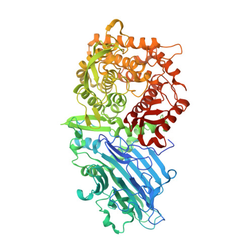
    4C1O, 4C1P

  • PubMed Abstract: 

    Geobacillus thermoglucosidasius is a thermophilic bacterium that is able to ferment both C6 and C5 sugars to produce ethanol. During growth on hemicellulose biomass, an intracellular β-xylosidase catalyses the hydrolysis of xylo-oligosaccharides to the monosaccharide xylose, which can then enter the pathways of central metabolism. The gene encoding a G. thermoglucosidasius β-xylosidase belonging to CAZy glycoside hydrolase family GH52 has been cloned and expressed in Escherichia coli. The recombinant enzyme has been characterized and a high-resolution (1.7 Å) crystal structure has been determined, resulting in the first reported structure of a GH52 family member. A lower resolution (2.6 Å) structure of the enzyme-substrate complex shows the positioning of the xylobiose substrate to be consistent with the proposed retaining mechanism of the family; additionally, the deep cleft of the active-site pocket, plus the proximity of the neighbouring subunit, afford an explanation for the lack of catalytic activity towards the polymer xylan. Whilst the fold of the G. thermoglucosidasius β-xylosidase is completely different from xylosidases in other CAZy families, the enzyme surprisingly shares structural similarities with other glycoside hydrolases, despite having no more than 13% sequence identity.


  • Organizational Affiliation
    • Centre for Extremophile Research, Department of Biology and Biochemistry, University of Bath, Claverton Down, Bath BA2 7AY, England.

Macromolecule Content 

  • Total Structure Weight: 82.61 kDa 
  • Atom Count: 6,007 
  • Modeled Residue Count: 703 
  • Deposited Residue Count: 727 
  • Unique protein chains: 1

Macromolecules

Find similar proteins by:|  3D Structure
Entity ID: 1
MoleculeChains  Sequence LengthOrganismDetailsImage
BETA-XYLOSIDASE727Parageobacillus thermoglucosidasiusMutation(s): 0 
EC: 3.2.1.37
UniProt
Find proteins for A0A067XG64 (Parageobacillus thermoglucosidasius)
Explore A0A067XG64 
Go to UniProtKB:  A0A067XG64
Entity Groups
Sequence Clusters30% Identity50% Identity70% Identity90% Identity95% Identity100% Identity
UniProt GroupA0A067XG64
Sequence Annotations
Expand
Reference Sequence

Oligosaccharides

Help  
Entity ID: 2
MoleculeChains Length2D Diagram GlycosylationD Interactions
beta-D-xylopyranose-(1-4)-beta-D-xylopyranose
B
2N/A
Glycosylation Resources
GlyTouCan: G87728WL
GlyCosmos: G87728WL

Biologically Interesting Molecules (External Reference) 

1 Unique

Experimental Data & Validation

Experimental Data

  • Method: X-RAY DIFFRACTION
  • Resolution: 2.63 Å
  • R-Value Free:  0.238 (Depositor), 0.239 (DCC) 
  • R-Value Work:  0.182 (Depositor), 0.183 (DCC) 
  • R-Value Observed: 0.185 (Depositor) 
Space Group: C 2 2 21
Unit Cell:
Length ( Å )Angle ( ˚ )
a = 73.369α = 90
b = 105.335β = 90
c = 194.834γ = 90
Software Package:
Software NamePurpose
PHENIXrefinement
HKL-2000data reduction
SCALEPACKdata scaling
REFMACphasing

Structure Validation

View Full Validation Report


Entry History 

Deposition Data

Revision History  (Full details and data files)

  • Version 1.0: 2014-05-14
    Type: Initial release
  • Version 1.1: 2014-05-21
    Changes: Database references
  • Version 1.2: 2019-05-08
    Changes: Data collection, Experimental preparation, Other
  • Version 2.0: 2020-07-29
    Type: Remediation
    Reason: Carbohydrate remediation
    Changes: Atomic model, Data collection, Derived calculations, Non-polymer description, Other, Structure summary
  • Version 2.1: 2024-05-01
    Changes: Data collection, Database references, Refinement description, Structure summary