4C0K | pdb_00004c0k

Crystal structure of Drosophila Miro EF hand and cGTPase domains bound to one calcium ion (Ca-MiroS)


Experimental Data Snapshot

  • Method: X-RAY DIFFRACTION
  • Resolution: 2.80 Å
  • R-Value Free: 
    0.242 (Depositor), 0.244 (DCC) 
  • R-Value Work: 
    0.207 (Depositor), 0.211 (DCC) 
  • R-Value Observed: 
    0.211 (Depositor) 

Starting Model: experimental
View more details

wwPDB Validation 3D Report Full Report

Validation slider image for 4C0K

This is version 2.1 of the entry. See complete history

Literature

Structural Coupling of the EF Hand and C-Terminal Gtpase Domains in the Mitochondrial Protein Miro.

Klosowiak, J.L.Focia, P.J.Chakravarthy, S.Landahl, E.C.Freymann, D.M.Rice, S.E.

(2013) EMBO Rep 14: 968

  • DOI: https://doi.org/10.1038/embor.2013.151
  • Primary Citation Related Structures: 
    4C0J, 4C0K, 4C0L

  • PubMed Abstract: 

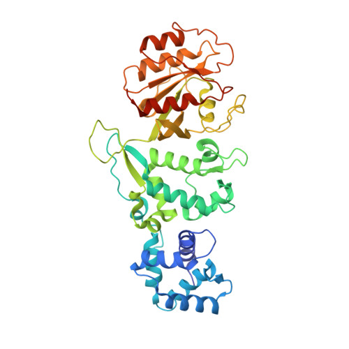
    Miro is a highly conserved calcium-binding GTPase at the regulatory nexus of mitochondrial transport and autophagy. Here we present crystal structures comprising the tandem EF hand and carboxy terminal GTPase (cGTPase) domains of Drosophila Miro. The structures reveal two previously unidentified 'hidden' EF hands, each paired with a canonical EF hand. Each EF hand pair is bound to a helix that structurally mimics an EF hand ligand. A key nucleotide-sensing element and a Pink1 phosphorylation site both lie within an extensive EF hand-cGTPase interface. Our results indicate structural mechanisms for calcium, nucleotide and phosphorylation-dependent regulation of mitochondrial function by Miro.


  • Organizational Affiliation
    • Department of Cell and Molecular Biology, Feinberg School of Medicine, Northwestern University, 303 East Chicago Avenue, Chicago, Illinois 60611, USA.

Macromolecule Content 

  • Total Structure Weight: 49.68 kDa 
  • Atom Count: 3,325 
  • Modeled Residue Count: 404 
  • Deposited Residue Count: 423 
  • Unique protein chains: 1

Macromolecules

Find similar proteins by:|  3D Structure
Entity ID: 1
MoleculeChains  Sequence LengthOrganismDetailsImage
MITOCHONDRIAL RHO GTPASE423Drosophila melanogasterMutation(s): 0 
EC: 3.6.5
UniProt
Find proteins for Q8IMX7 (Drosophila melanogaster)
Explore Q8IMX7 
Go to UniProtKB:  Q8IMX7
Entity Groups
Sequence Clusters30% Identity50% Identity70% Identity90% Identity95% Identity100% Identity
UniProt GroupQ8IMX7
Sequence Annotations
Expand
Reference Sequence

Small Molecules

Ligands 5 Unique
IDChains Name / Formula / InChI Key2D Diagram3D Interactions
HSE

Query on HSE


Download:Ideal Coordinates CCD File
B [auth A]L-HOMOSERINE
C4 H9 N O3
UKAUYVFTDYCKQA-VKHMYHEASA-N
SO4

Query on SO4


Download:Ideal Coordinates CCD File
D [auth A],
E [auth A],
F [auth A],
G [auth A]
SULFATE ION
O4 S
QAOWNCQODCNURD-UHFFFAOYSA-L
CA

Query on CA


Download:Ideal Coordinates CCD File
I [auth A]CALCIUM ION
Ca
BHPQYMZQTOCNFJ-UHFFFAOYSA-N
NA

Query on NA


Download:Ideal Coordinates CCD File
H [auth A]SODIUM ION
Na
FKNQFGJONOIPTF-UHFFFAOYSA-N
UNX

Query on UNX


Download:Ideal Coordinates CCD File
C [auth A]UNKNOWN ATOM OR ION
X

Experimental Data & Validation

Experimental Data

  • Method: X-RAY DIFFRACTION
  • Resolution: 2.80 Å
  • R-Value Free:  0.242 (Depositor), 0.244 (DCC) 
  • R-Value Work:  0.207 (Depositor), 0.211 (DCC) 
  • R-Value Observed: 0.211 (Depositor) 
Space Group: P 32 2 1
Unit Cell:
Length ( Å )Angle ( ˚ )
a = 82.24α = 90
b = 82.24β = 90
c = 156.441γ = 120
Software Package:
Software NamePurpose
PHENIXrefinement
MOSFLMdata reduction
Aimlessdata scaling
PHENIXphasing

Structure Validation

View Full Validation Report


Entry History 

Deposition Data

Revision History  (Full details and data files)

  • Version 1.0: 2013-10-09
    Type: Initial release
  • Version 1.1: 2013-11-13
    Changes: Database references
  • Version 2.0: 2023-11-15
    Changes: Atomic model, Data collection, Database references, Derived calculations, Other
  • Version 2.1: 2024-05-01
    Changes: Refinement description