4C09 | pdb_00004c09

Crystal structure of the metallo-beta-lactamase BCII


Experimental Data Snapshot

  • Method: X-RAY DIFFRACTION
  • Resolution: 1.20 Å
  • R-Value Free: 
    0.139 (Depositor), 0.151 (DCC) 
  • R-Value Work: 
    0.119 (Depositor), 0.133 (DCC) 
  • R-Value Observed: 
    0.119 (Depositor) 

Starting Model: experimental
View more details

wwPDB Validation 3D Report Full Report

Validation slider image for 4C09

This is version 1.5 of the entry. See complete history

Literature

Structural Basis of Metallo-beta-Lactamase Inhibition by Captopril Stereoisomers.

Brem, J.van Berkel, S.S.Zollman, D.Lee, S.Y.Gileadi, O.McHugh, P.J.Walsh, T.R.McDonough, M.A.Schofield, C.J.

(2015) Antimicrob Agents Chemother 60: 142-150

  • DOI: https://doi.org/10.1128/AAC.01335-15
  • Primary Citation Related Structures: 
    4BZ3, 4C09, 4C1C, 4C1D, 4C1E, 4C1F, 4C1G, 4C1H

  • PubMed Abstract: 

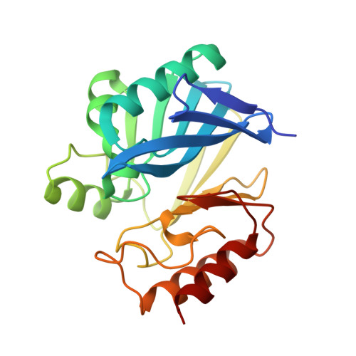
    β-Lactams are the most successful antibacterials, but their effectiveness is threatened by resistance, most importantly by production of serine- and metallo-β-lactamases (MBLs). MBLs are of increasing concern because they catalyze the hydrolysis of almost all β-lactam antibiotics, including recent-generation carbapenems. Clinically useful serine-β-lactamase inhibitors have been developed, but such inhibitors are not available for MBLs. l-Captopril, which is used to treat hypertension via angiotensin-converting enzyme inhibition, has been reported to inhibit MBLs by chelating the active site zinc ions via its thiol(ate). We report systematic studies on B1 MBL inhibition by all four captopril stereoisomers. High-resolution crystal structures of three MBLs (IMP-1, BcII, and VIM-2) in complex with either the l- or d-captopril stereoisomer reveal correlations between the binding mode and inhibition potency. The results will be useful in the design of MBL inhibitors with the breadth of selectivity required for clinical application against carbapenem-resistant Enterobacteriaceae and other organisms causing MBL-mediated resistant infections.


  • Organizational Affiliation
    • Department of Chemistry, University of Oxford, Oxford, United Kingdom.

Macromolecule Content 

  • Total Structure Weight: 25.41 kDa 
  • Atom Count: 2,124 
  • Modeled Residue Count: 223 
  • Deposited Residue Count: 227 
  • Unique protein chains: 1

Macromolecules

Find similar proteins by:|  3D Structure
Entity ID: 1
MoleculeChains  Sequence LengthOrganismDetailsImage
BETA-LACTAMASE 2227Bacillus cereusMutation(s): 0 
EC: 3.5.2.6
UniProt
Find proteins for P04190 (Bacillus cereus)
Explore P04190 
Go to UniProtKB:  P04190
Entity Groups
Sequence Clusters30% Identity50% Identity70% Identity90% Identity95% Identity100% Identity
UniProt GroupP04190
Sequence Annotations
Expand
Reference Sequence

Experimental Data & Validation

Experimental Data

  • Method: X-RAY DIFFRACTION
  • Resolution: 1.20 Å
  • R-Value Free:  0.139 (Depositor), 0.151 (DCC) 
  • R-Value Work:  0.119 (Depositor), 0.133 (DCC) 
  • R-Value Observed: 0.119 (Depositor) 
Space Group: C 1 2 1
Unit Cell:
Length ( Å )Angle ( ˚ )
a = 53.3α = 90
b = 61.97β = 93.04
c = 69.46γ = 90
Software Package:
Software NamePurpose
PHENIXrefinement
XDSdata reduction
XSCALEdata scaling
PHASERphasing

Structure Validation

View Full Validation Report


Entry History 

Deposition Data

Revision History  (Full details and data files)

  • Version 1.0: 2014-08-20
    Type: Initial release
  • Version 1.1: 2014-09-17
    Changes: Other, Structure summary
  • Version 1.2: 2015-11-25
    Changes: Database references
  • Version 1.3: 2016-01-13
    Changes: Database references
  • Version 1.4: 2018-02-21
    Changes: Database references, Structure summary
  • Version 1.5: 2023-12-20
    Changes: Data collection, Database references, Derived calculations, Other, Refinement description